Moewis Philippe, Duda Georg N, Jung Tobias, Heller Markus O, Boeth Heide, Kaptein Bart, Taylor William R
Julius Wolff Institute, Charité-Universitätsmedizin Berlin, Berlin, Germany.
Knee Surgery and Sports Traumatology, Center for Musculoskeletal Surgery, Charité-Universitätsmedizin Berlin, Berlin, Germany.
PLoS One. 2016 Jul 28;11(7):e0159600. doi: 10.1371/journal.pone.0159600. eCollection 2016.
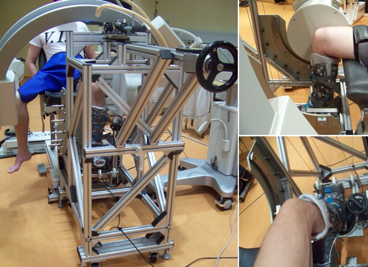
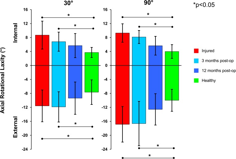
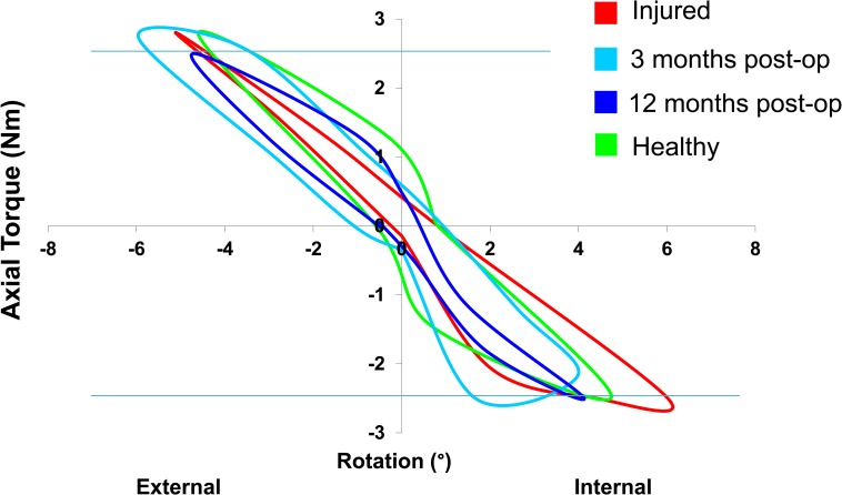
While the anterior cruciate ligament (ACL) is considered one of the most important ligaments for providing knee joint stability, its influence on rotational laxity is not fully understood and its role in resisting rotation at different flexion angles in vivo remains unknown. In this prospective study, we investigated the relationship between in vivo passive axial rotational laxity and knee flexion angle, as well as how they were altered with ACL injury and reconstruction. A rotometer device was developed to assess knee joint rotational laxity under controlled passive testing. An axial torque of ±2.5Nm was applied to the knee while synchronised fluoroscopic images of the tibia and femur allowed axial rotation of the bones to be accurately determined. Passive rotational laxity tests were completed in 9 patients with an untreated ACL injury and compared to measurements at 3 and 12 months after anatomical single bundle ACL reconstruction, as well as to the contralateral controls. Significant differences in rotational laxity were found between the injured and the healthy contralateral knees with internal rotation values of 8.7°±4.0° and 3.7°±1.4° (p = 0.003) at 30° of flexion and 9.3°±2.6° and 4.0°±2.0° (p = 0.001) at 90° respectively. After 3 months, the rotational laxity remained similar to the injured condition, and significantly different to the healthy knees. However, after 12 months, a considerable reduction of rotational laxity was observed towards the levels of the contralateral controls. The significantly greater laxity observed at both knee flexion angles after 3 months (but not at 12 months), suggests an initial lack of post-operative rotational stability, possibly due to reduced mechanical properties or fixation stability of the graft tissue. After 12 months, reduced levels of rotational laxity compared with the injured and 3 month conditions, both internally and externally, suggests progressive rotational stability of the reconstruction with time.
虽然前交叉韧带(ACL)被认为是提供膝关节稳定性的最重要韧带之一,但其对旋转松弛的影响尚未完全了解,其在体内不同屈曲角度抵抗旋转的作用仍不清楚。在这项前瞻性研究中,我们调查了体内被动轴向旋转松弛与膝关节屈曲角度之间的关系,以及它们如何因ACL损伤和重建而改变。开发了一种旋转计装置,以在受控的被动测试下评估膝关节旋转松弛。在向膝关节施加±2.5 Nm的轴向扭矩时,通过同步的胫骨和股骨荧光透视图像,可以准确确定骨骼的轴向旋转。对9例未经治疗的ACL损伤患者进行了被动旋转松弛测试,并与解剖单束ACL重建后3个月和12个月的测量结果以及对侧对照进行了比较。在受伤膝关节和健康对侧膝关节之间发现旋转松弛存在显著差异,屈曲30°时内旋值分别为8.7°±4.0°和3.7°±1.4°(p = 0.003),屈曲90°时分别为9.3°±2.6°和4.0°±2.0°(p = 0.001)。3个月后,旋转松弛仍与受伤状态相似,与健康膝关节有显著差异。然而,12个月后,观察到旋转松弛显著降低,接近对侧对照水平。3个月后(但12个月时未观察到)在两个膝关节屈曲角度均观察到的显著更大松弛,表明术后最初缺乏旋转稳定性,可能是由于移植物组织的力学性能或固定稳定性降低。12个月后,与受伤和3个月时相比,内旋和外旋的旋转松弛水平均降低,表明重建随时间逐渐获得旋转稳定性。