• 文献检索
  • 文档翻译
  • 深度研究
  • 学术资讯
  • Suppr Zotero 插件Zotero 插件
  • 邀请有礼
  • 套餐&价格
  • 历史记录
应用&插件
Suppr Zotero 插件Zotero 插件浏览器插件Mac 客户端Windows 客户端微信小程序
定价
高级版会员购买积分包购买API积分包
服务
文献检索文档翻译深度研究API 文档MCP 服务
关于我们
关于 Suppr公司介绍联系我们用户协议隐私条款
关注我们

Suppr 超能文献

核心技术专利:CN118964589B侵权必究
粤ICP备2023148730 号-1Suppr @ 2026

文献检索

告别复杂PubMed语法,用中文像聊天一样搜索,搜遍4000万医学文献。AI智能推荐,让科研检索更轻松。

立即免费搜索

文件翻译

保留排版,准确专业,支持PDF/Word/PPT等文件格式,支持 12+语言互译。

免费翻译文档

深度研究

AI帮你快速写综述,25分钟生成高质量综述,智能提取关键信息,辅助科研写作。

立即免费体验

T1-VISTA辅助诊断和随访小脑后下动脉夹层并发缺血性卒中:两例报告

Diagnosis and follow-up of posterior inferior cerebellar artery dissection complicated with ischemic stroke assisted by T1-VISTA: a report of two cases.

作者信息

Ishitsuka Koji, Sakaki Yusuke, Sakai Shota, Uwatoko Takeshi, Aibe Hitoshi, Ago Tetsuro, Kitazono Takanari, Sugimori Hiroshi

机构信息

Department of Cerebrovascular Medicine, Saga Medical Center Koseikan, Kase-machi Nakabaru 400, Saga, 840-8571, Japan.

Department of Cerebrovascular Disease, Japanese Fukuoka Red Cross Hospital, Fukuoka, 815-8555, Japan.

出版信息

BMC Neurol. 2016 Jul 29;16:121. doi: 10.1186/s12883-016-0637-9.

DOI:10.1186/s12883-016-0637-9
PMID:27474010
原文链接:https://pmc.ncbi.nlm.nih.gov/articles/PMC4966802/
Abstract

BACKGROUND

Volume isotropic turbo spin-echo acquisition (VISTA) is a new method similar to the 3D black-blood imaging method that enables visualization of a intramural hematoma. T1-VISTA has recently been applied in the diagnosis of intracranial arterial dissection. However, the identification of an intramural hematoma in posterior inferior cerebellar dissection (PICA-D) by T1-VISTA has only rarely been reported.

CASE PRESENTATION

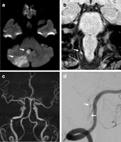
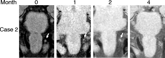
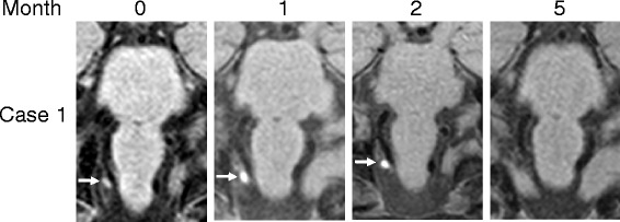
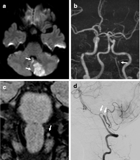
We herein report two patients who suffered from PICA-D complicated with ischemic stroke. Initial magnetic resonance arteriography was not informative, however, T1-VISTA depicted high-intensity signal areas suggesting an intramural hematoma of PICA-D in both cases. The high-intensity signal areas gradually reduced and finally disappeared at 4 months and 5 months after the onset, respectively.

CONCLUSION

Our cases demonstrate that T1-VISTA was able to assist in the diagnosis and follow-up of PICA-D.

摘要

背景

容积各向同性涡轮自旋回波采集(VISTA)是一种类似于三维黑血成像方法的新方法,能够实现壁内血肿的可视化。T1-VISTA最近已应用于颅内动脉夹层的诊断。然而,通过T1-VISTA识别小脑后下动脉夹层(PICA-D)中的壁内血肿的报道很少。

病例报告

我们在此报告2例患有PICA-D并伴有缺血性卒中的患者。最初的磁共振血管造影未提供有用信息,然而,T1-VISTA在2例病例中均显示出高强度信号区域,提示PICA-D的壁内血肿。高强度信号区域分别在发病后4个月和5个月逐渐缩小并最终消失。

结论

我们的病例表明,T1-VISTA能够辅助PICA-D的诊断和随访。

https://cdn.ncbi.nlm.nih.gov/pmc/blobs/d887/4966802/b39c859ec227/12883_2016_637_Fig4_HTML.jpg
https://cdn.ncbi.nlm.nih.gov/pmc/blobs/d887/4966802/b9d54e250cbc/12883_2016_637_Fig1_HTML.jpg
https://cdn.ncbi.nlm.nih.gov/pmc/blobs/d887/4966802/57e6bb506ae8/12883_2016_637_Fig2_HTML.jpg
https://cdn.ncbi.nlm.nih.gov/pmc/blobs/d887/4966802/a01b992acb15/12883_2016_637_Fig3_HTML.jpg
https://cdn.ncbi.nlm.nih.gov/pmc/blobs/d887/4966802/b39c859ec227/12883_2016_637_Fig4_HTML.jpg

相似文献

1
Diagnosis and follow-up of posterior inferior cerebellar artery dissection complicated with ischemic stroke assisted by T1-VISTA: a report of two cases.T1-VISTA辅助诊断和随访小脑后下动脉夹层并发缺血性卒中:两例报告
BMC Neurol. 2016 Jul 29;16:121. doi: 10.1186/s12883-016-0637-9.
2
Intimal flap detected by three-dimensional curved multiplanar reconstruction image in isolated posterior inferior cerebellar artery dissection: a report of two cases.三维曲面多平面重建图像检测孤立性小脑后下动脉夹层中的内膜瓣:两例报告
BMC Neurol. 2019 Apr 27;19(1):74. doi: 10.1186/s12883-019-1309-3.
3
Utility of T1- and T2-Weighted High-Resolution Vessel Wall Imaging for the Diagnosis and Follow Up of Isolated Posterior Inferior Cerebellar Artery Dissection with Ischemic Stroke: Report of 4 Cases and Review of the Literature.T1加权和T2加权高分辨率血管壁成像在孤立性小脑后下动脉夹层伴缺血性卒中诊断及随访中的应用:4例报告并文献复习
J Stroke Cerebrovasc Dis. 2017 Nov;26(11):2645-2651. doi: 10.1016/j.jstrokecerebrovasdis.2017.06.038. Epub 2017 Aug 30.
4
Evaluation of luminal and vessel wall abnormalities in subacute and other stages of intracranial vertebrobasilar artery dissections using the volume isotropic turbo-spin-echo acquisition (VISTA) sequence: a preliminary study.应用容积各向同性翻转回波序列(VISTA)评价颅内外椎动脉夹层亚急性期及其他时期管腔及血管壁异常:初步研究
J Neuroradiol. 2013 Mar;40(1):19-28. doi: 10.1016/j.neurad.2012.02.005. Epub 2012 May 25.
5
3D T1-weighted black blood sequence at 3.0 Tesla for the diagnosis of cervical artery dissection.3.0T 磁共振 3D T1-weighted 黑血序列在诊断颈动脉夹层中的应用
Stroke Vasc Neurol. 2016 Oct 25;1(3):140-146. doi: 10.1136/svn-2016-000028. eCollection 2016 Sep.
6
Evaluating intracranial artery dissection by using three-dimensional simultaneous non-contrast angiography and intra-plaque hemorrhage high-resolution magnetic resonance imaging: a retrospective study.利用三维同步非对比血管造影和斑块内出血高分辨率磁共振成像评估颅内动脉夹层:一项回顾性研究。
Acta Radiol. 2022 Mar;63(3):401-409. doi: 10.1177/0284185121992235. Epub 2021 Feb 18.
7
Added Value of 3D Proton-Density Weighted Images in Diagnosis of Intracranial Arterial Dissection.三维质子密度加权成像在颅内动脉夹层诊断中的附加价值
PLoS One. 2016 Nov 23;11(11):e0166929. doi: 10.1371/journal.pone.0166929. eCollection 2016.
8
Do characteristics of dissection differ between the posterior inferior cerebellar artery and the vertebral artery?小脑后下动脉和椎动脉的夹层特征有差异吗?
J Stroke Cerebrovasc Dis. 2014 Nov-Dec;23(10):2857-2861. doi: 10.1016/j.jstrokecerebrovasdis.2014.07.013. Epub 2014 Oct 3.
9
Intracranial Cerebral Artery Dissection of Anterior Circulation as a Cause of Convexity Subarachnoid Hemorrhage.前循环颅内脑动脉夹层作为凸面蛛网膜下腔出血的病因
Cerebrovasc Dis. 2015;40(1-2):45-51. doi: 10.1159/000430945. Epub 2015 Jun 16.
10
Remote distal outflow occlusion: a novel treatment option for complex dissecting aneurysms of the posterior inferior cerebellar artery. Report of 3 cases.远端流出道闭塞:一种治疗小脑后下动脉复杂夹层动脉瘤的新方法。附3例报告。
J Neurosurg. 2009 Jul;111(1):78-83. doi: 10.3171/2009.1.JNS081250.

引用本文的文献

1
Traumatic dissection of the anterior cerebral artery secondary to a rugby related impact: A case report with emphasis on the usefulness of T1-VISTA.橄榄球相关撞击继发大脑前动脉创伤性夹层:一例报告并强调T1-VISTA的实用性
Surg Neurol Int. 2023 Feb 3;14:43. doi: 10.25259/SNI_1082_2022. eCollection 2023.
2
Qualitative Assessment and Reporting Quality of Intracranial Vessel Wall MR Imaging Studies: A Systematic Review.颅内血管壁磁共振成像研究的定性评估和报告质量:系统评价。
AJNR Am J Neuroradiol. 2019 Dec;40(12):2025-2032. doi: 10.3174/ajnr.A6317. Epub 2019 Nov 14.
3
Intimal flap detected by three-dimensional curved multiplanar reconstruction image in isolated posterior inferior cerebellar artery dissection: a report of two cases.

本文引用的文献

1
Intracranial and extracranial arterial dissection presenting with ischemic stroke: Lesion location and stroke mechanism.以缺血性卒中为表现的颅内和颅外动脉夹层:病变部位与卒中机制
J Neurol Sci. 2015 Nov 15;358(1-2):371-6. doi: 10.1016/j.jns.2015.09.368. Epub 2015 Sep 28.
2
3D fat-saturated T1 SPACE sequence for the diagnosis of cervical artery dissection.3D 脂肪饱和 T1 SPACE 序列在诊断颈动脉夹层中的应用。
Neuroradiology. 2013 May;55(5):595-602. doi: 10.1007/s00234-013-1141-1. Epub 2013 Jan 25.
3
3D fast spin-echo T1 black-blood imaging for the diagnosis of cervical artery dissection.
三维曲面多平面重建图像检测孤立性小脑后下动脉夹层中的内膜瓣:两例报告
BMC Neurol. 2019 Apr 27;19(1):74. doi: 10.1186/s12883-019-1309-3.
4
Clinical Characteristics of Cerebellar Infarction Due to Arterial Dissection.动脉夹层所致小脑梗死的临床特征
Asian J Neurosurg. 2018 Oct-Dec;13(4):995-1000. doi: 10.4103/ajns.AJNS_373_16.
5
Utility of the variable flip angle 3D fast-spin echo (isoFSE) sequence on 3T MR for diagnosing vertebrobasilar artery dissection.3T磁共振成像中可变翻转角三维快速自旋回波(isoFSE)序列在诊断椎基底动脉夹层中的应用价值。
Radiol Phys Technol. 2018 Jun;11(2):228-234. doi: 10.1007/s12194-018-0460-7. Epub 2018 Apr 25.
3D 快速自旋回波 T1 黑血成像在诊断颈内动脉夹层中的应用。
AJNR Am J Neuroradiol. 2013 Sep;34(9):E103-6. doi: 10.3174/ajnr.A3261. Epub 2012 Oct 11.
4
Evaluation of luminal and vessel wall abnormalities in subacute and other stages of intracranial vertebrobasilar artery dissections using the volume isotropic turbo-spin-echo acquisition (VISTA) sequence: a preliminary study.应用容积各向同性翻转回波序列(VISTA)评价颅内外椎动脉夹层亚急性期及其他时期管腔及血管壁异常:初步研究
J Neuroradiol. 2013 Mar;40(1):19-28. doi: 10.1016/j.neurad.2012.02.005. Epub 2012 May 25.
5
The new MRI modalities "BPAS and VISTA" for the diagnosis of VA dissection.
Acta Neurochir Suppl. 2011;112:59-65. doi: 10.1007/978-3-7091-0661-7_11.
6
Traumatic intracranial dissection: mural hematoma on high-resolution MRI.创伤性颅内夹层动脉瘤:高分辨率磁共振成像显示壁间血肿
J Neuroradiol. 2010 May;37(2):136-7. doi: 10.1016/j.neurad.2009.07.002. Epub 2009 Sep 18.
7
Vertebral artery dissection with a normal-appearing lumen at multisection CT angiography: the importance of identifying wall hematoma.多层CT血管造影显示管腔正常的椎动脉夹层:识别壁内血肿的重要性
AJNR Am J Neuroradiol. 2009 Apr;30(4):787-92. doi: 10.3174/ajnr.A1455. Epub 2009 Jan 22.
8
Surface appearance of the vertebrobasilar artery revealed on basiparallel anatomic scanning (BPAS)-MR imaging: its role for brain MR examination.基底平行解剖扫描(BPAS)-磁共振成像显示的椎基底动脉表面形态:其在脑部磁共振检查中的作用
AJNR Am J Neuroradiol. 2005 Nov-Dec;26(10):2508-13.
9
Isolated dissections and dissecting aneurysms of the posterior inferior cerebellar artery: topic and literature review.小脑后下动脉孤立性夹层及夹层动脉瘤:主题与文献综述
Neurosurg Rev. 2003 Jul;26(3):180-7. doi: 10.1007/s10143-002-0231-z. Epub 2002 Sep 4.
10
Spontaneous dissection of the carotid and vertebral arteries.颈动脉和椎动脉自发性夹层
N Engl J Med. 2001 Mar 22;344(12):898-906. doi: 10.1056/NEJM200103223441206.