Wang Peng, Yao Lan, Zhou Li-Li, Liu Yuan-Shan, Chen Ming-di, Wu Hai-Dong, Chang Rui-Ming, Li Yi, Zhou Ming-Gen, Fang Xiang-Shao, Yu Tao, Jiang Long-Yuan, Huang Zi-Tong
1. Department of Emergency Medicine, Sun Yat-sen Memorial Hospital of Sun Yat-sen University, Guangzhou, China; 2. Institute of Cardiopulmonary Cerebral Resuscitation, Sun Yat-sen University, Guangzhou, China.
1. Department of Emergency Medicine, Sun Yat-sen Memorial Hospital of Sun Yat-sen University, Guangzhou, China; 2. Institute of Cardiopulmonary Cerebral Resuscitation, Sun Yat-sen University, Guangzhou, China; 3. Department of Emergency Medicine, The fifth affiliated hospital, Sun Yat-sen University, Zhuhai, China.
Int J Biol Sci. 2016 Jul 17;12(8):1000-9. doi: 10.7150/ijbs.13222. eCollection 2016.
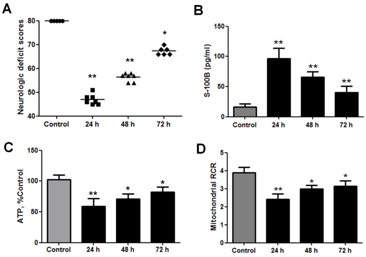
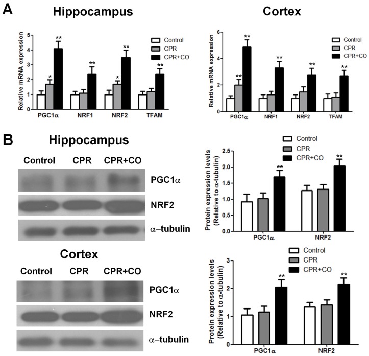
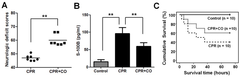
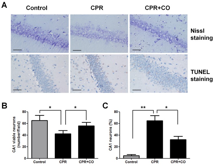
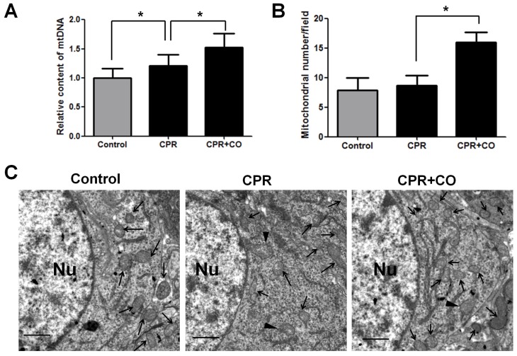
Mitochondrial dysfunction contributes to brain injury following global cerebral ischemia after cardiac arrest. Carbon monoxide treatment has shown potent cytoprotective effects in ischemia/reperfusion injury. This study aimed to investigate the effects of carbon monoxide-releasing molecules on brain mitochondrial dysfunction and brain injury following resuscitation after cardiac arrest in rats. A rat model of cardiac arrest was established by asphyxia. The animals were randomly divided into the following 3 groups: cardiac arrest and resuscitation group, cardiac arrest and resuscitation plus carbon monoxide intervention group, and sham control group (no cardiac arrest). After the return of spontaneous circulation, neurologic deficit scores (NDS) and S-100B levels were significantly decreased at 24, 48, and 72 h, but carbon monoxide treatment improved the NDS and S-100B levels at 24 h and the 3-day survival rates of the rats. This treatment also decreased the number of damaged neurons in the hippocampus CA1 area and increased the brain mitochondrial activity. In addition, it increased mitochondrial biogenesis by increasing the expression of biogenesis factors including peroxisome proliferator-activated receptor-γ coactivator-1α, nuclear respiratory factor-1, nuclear respiratory factor-2 and mitochondrial transcription factor A. Thus, this study showed that carbon monoxide treatment alleviated brain injury after cardiac arrest in rats by increased brain mitochondrial biogenesis.
线粒体功能障碍会导致心脏骤停后全脑缺血性脑损伤。一氧化碳治疗已显示出对缺血/再灌注损伤具有强大的细胞保护作用。本研究旨在探讨一氧化碳释放分子对大鼠心脏骤停复苏后脑线粒体功能障碍和脑损伤的影响。通过窒息建立大鼠心脏骤停模型。将动物随机分为以下3组:心脏骤停与复苏组、心脏骤停与复苏加一氧化碳干预组和假手术对照组(无心脏骤停)。自主循环恢复后,神经功能缺损评分(NDS)和S-100B水平在24、48和72小时时显著降低,但一氧化碳治疗改善了24小时时的NDS和S-100B水平以及大鼠的3天生存率。该治疗还减少了海马CA1区受损神经元的数量并增加了脑线粒体活性。此外,它通过增加包括过氧化物酶体增殖物激活受体-γ共激活因子-1α、核呼吸因子-1、核呼吸因子-2和线粒体转录因子A在内的生物发生因子的表达来增加线粒体生物发生。因此,本研究表明一氧化碳治疗通过增加脑线粒体生物发生减轻了大鼠心脏骤停后的脑损伤。