• 文献检索
  • 文档翻译
  • 深度研究
  • 学术资讯
  • Suppr Zotero 插件Zotero 插件
  • 邀请有礼
  • 套餐&价格
  • 历史记录
应用&插件
Suppr Zotero 插件Zotero 插件浏览器插件Mac 客户端Windows 客户端微信小程序
定价
高级版会员购买积分包购买API积分包
服务
文献检索文档翻译深度研究API 文档MCP 服务
关于我们
关于 Suppr公司介绍联系我们用户协议隐私条款
关注我们

Suppr 超能文献

核心技术专利:CN118964589B侵权必究
粤ICP备2023148730 号-1Suppr @ 2026

文献检索

告别复杂PubMed语法,用中文像聊天一样搜索,搜遍4000万医学文献。AI智能推荐,让科研检索更轻松。

立即免费搜索

文件翻译

保留排版,准确专业,支持PDF/Word/PPT等文件格式,支持 12+语言互译。

免费翻译文档

深度研究

AI帮你快速写综述,25分钟生成高质量综述,智能提取关键信息,辅助科研写作。

立即免费体验

一种用于对III级胶质瘤活检标本中全脑磁共振波谱成像与组织学进行客观比较的系统流程。

A systematic pipeline for the objective comparison of whole-brain spectroscopic MRI with histology in biopsy specimens from grade III glioma.

作者信息

Cordova J Scott, Gurbani Saumya S, Olson Jeffrey J, Liang Zhongxing, Cooper Lee A D, Shu Hui-Kuo G, Schreibmann Eduard, Neill Stewart G, Hadjipanayis Constantinos G, Holder Chad A, Shim Hyunsuk

机构信息

Department of Radiology and Imaging Sciences, Emory University School of Medicine, Atlanta, GA.

Department of Radiology and Imaging Sciences, Emory University School of Medicine, Atlanta, GA; Department of Biomedical Engineering, Georgia Institute of Technology, Atlanta, GA.

出版信息

Tomography. 2016 Jun;2(2):106-116. doi: 10.18383/j.tom.2016.00136.

DOI:10.18383/j.tom.2016.00136
PMID:27489883
原文链接:https://pmc.ncbi.nlm.nih.gov/articles/PMC4968944/
Abstract

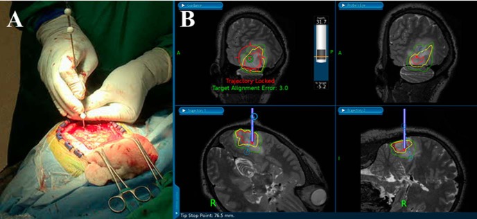
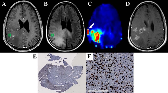
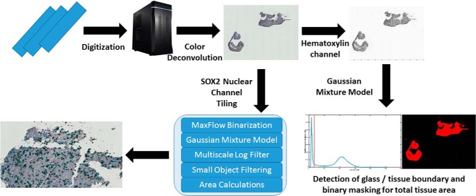
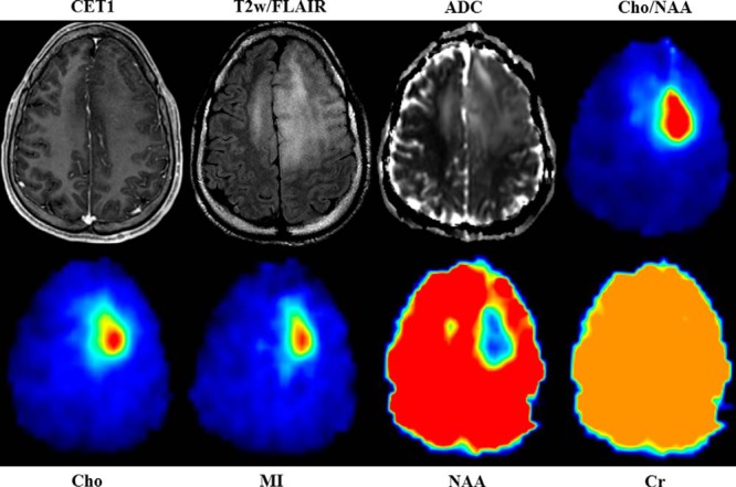
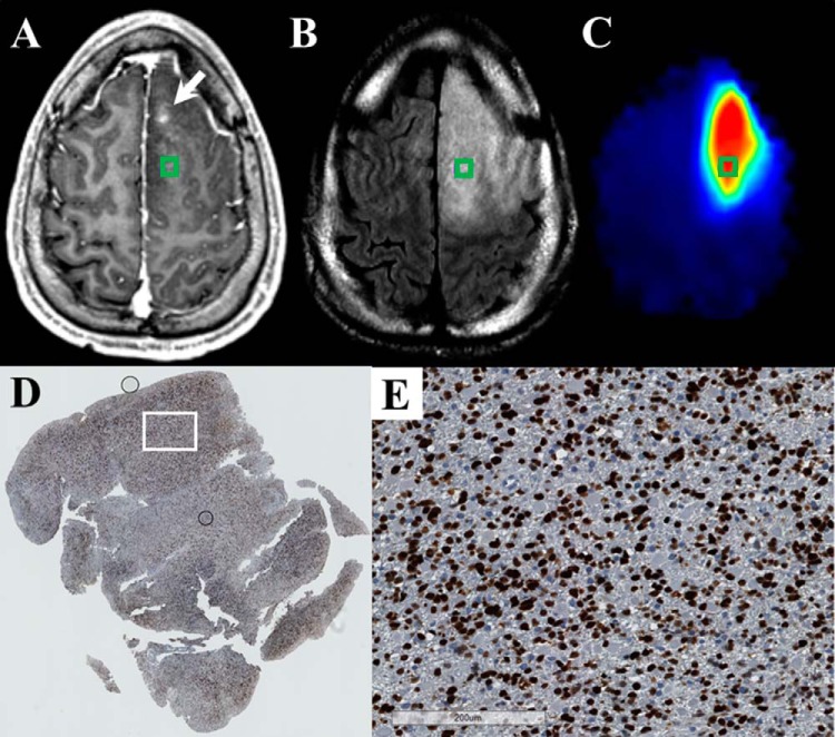
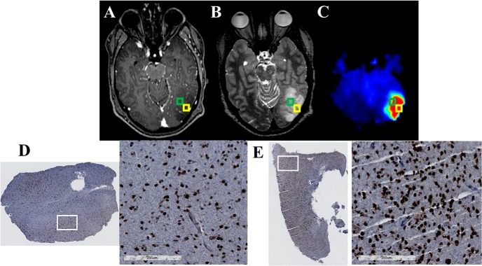
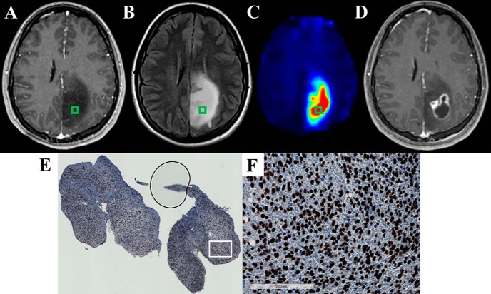
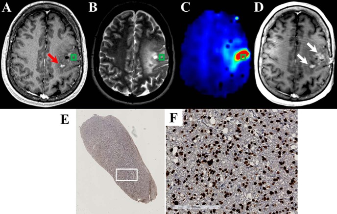
The diagnosis, prognosis, and management of patients with gliomas are largely dictated by the pathological analysis of tissue biopsied from a selected region within the lesion. However, due to the heterogeneous and infiltrative nature of gliomas, identifying the optimal region for biopsy with conventional magnetic resonance imaging (MRI) can be quite difficult. This is especially true for low grade gliomas, which often are non-enhancing tumors. To improve the management of patients with these tumors, the field of neuro-oncology requires an imaging modality that can specifically identify a tumor's most anaplastic/aggressive region(s) for biopsy targeting. The addition of metabolic mapping using spectroscopic MRI (sMRI) to supplement conventional MRI could improve biopsy targeting and, ultimately, diagnostic accuracy. Here, we describe a pipeline for the integration of state-of-the-art, high-resolution whole-brain 3D sMRI maps into a stereotactic neuronavigation system for guiding biopsies in gliomas with nonenhancing components. We also outline a machine-learning method for automated histology analysis that generates normalized, quantitative metrics describing tumor infiltration in immunohistochemically-stained tissue specimens. As a proof of concept, we describe the combination of these two techniques in a small cohort of grade III glioma patients. In this work, we aim to set forth a systematic pipeline to stimulate histopathology-image validation of advanced MRI techniques, such as sMRI.

摘要

胶质瘤患者的诊断、预后及治疗很大程度上取决于对病变内选定区域活检组织的病理分析。然而,由于胶质瘤具有异质性和浸润性,利用传统磁共振成像(MRI)确定活检的最佳区域可能相当困难。对于低级别胶质瘤尤其如此,这类肿瘤通常不强化。为改善这类肿瘤患者的治疗,神经肿瘤学领域需要一种成像方式,能够特异性地识别肿瘤最间变/侵袭性的区域以进行活检定位。添加使用磁共振波谱成像(sMRI)的代谢图谱以补充传统MRI,可改善活检定位,并最终提高诊断准确性。在此,我们描述了一种流程,用于将先进的高分辨率全脑三维sMRI图谱整合到立体定向神经导航系统中,以指导对含有非强化成分的胶质瘤进行活检。我们还概述了一种用于自动组织学分析的机器学习方法,该方法可生成描述免疫组织化学染色组织标本中肿瘤浸润情况的标准化定量指标。作为概念验证,我们在一小群III级胶质瘤患者中描述了这两种技术的结合。在这项工作中,我们旨在提出一个系统流程,以推动对诸如sMRI等先进MRI技术进行组织病理学图像验证。

https://cdn.ncbi.nlm.nih.gov/pmc/blobs/4041/6024420/3a5e217c01b2/tom0021600330008.jpg
https://cdn.ncbi.nlm.nih.gov/pmc/blobs/4041/6024420/ac148fdd6e4b/tom0021600330001.jpg
https://cdn.ncbi.nlm.nih.gov/pmc/blobs/4041/6024420/5e2cdf4a1681/tom0021600330002.jpg
https://cdn.ncbi.nlm.nih.gov/pmc/blobs/4041/6024420/0229b840438c/tom0021600330003.jpg
https://cdn.ncbi.nlm.nih.gov/pmc/blobs/4041/6024420/12ba5758ce91/tom0021600330004.jpg
https://cdn.ncbi.nlm.nih.gov/pmc/blobs/4041/6024420/2faf5300431d/tom0021600330005.jpg
https://cdn.ncbi.nlm.nih.gov/pmc/blobs/4041/6024420/9965db5d9025/tom0021600330006.jpg
https://cdn.ncbi.nlm.nih.gov/pmc/blobs/4041/6024420/fc93719dfbcd/tom0021600330007.jpg
https://cdn.ncbi.nlm.nih.gov/pmc/blobs/4041/6024420/3a5e217c01b2/tom0021600330008.jpg

相似文献

1
A systematic pipeline for the objective comparison of whole-brain spectroscopic MRI with histology in biopsy specimens from grade III glioma.一种用于对III级胶质瘤活检标本中全脑磁共振波谱成像与组织学进行客观比较的系统流程。
Tomography. 2016 Jun;2(2):106-116. doi: 10.18383/j.tom.2016.00136.
2
Biopsy targeting gliomas: do functional imaging techniques identify similar target areas?活检靶向胶质瘤:功能成像技术能否识别出相似的靶区?
Invest Radiol. 2010 Dec;45(12):755-68. doi: 10.1097/RLI.0b013e3181ec9db0.
3
Introduction of a standardized multimodality image protocol for navigation-guided surgery of suspected low-grade gliomas.用于疑似低级别胶质瘤导航引导手术的标准化多模态影像方案介绍。
Neurosurg Focus. 2015 Jan;38(1):E4. doi: 10.3171/2014.10.FOCUS14597.
4
Whole-brain spectroscopic MRI biomarkers identify infiltrating margins in glioblastoma patients.全脑磁共振波谱成像生物标志物可识别胶质母细胞瘤患者的浸润边缘。
Neuro Oncol. 2016 Aug;18(8):1180-9. doi: 10.1093/neuonc/now036. Epub 2016 Mar 15.
5
11C-Methionine positron emission tomography for preoperative evaluation of suggestive low-grade gliomas.11C-蛋氨酸正电子发射断层扫描用于提示性低级别胶质瘤的术前评估
Zentralbl Neurochir. 2007 Feb;68(1):19-23. doi: 10.1055/s-2007-970601.
6
Accuracy of frameless and frame-based image-guided stereotactic brain biopsy in the diagnosis of glioma: comparison of biopsy and open resection specimen.无框架和基于框架的影像引导立体定向脑活检在胶质瘤诊断中的准确性:活检与开放切除标本的比较
Neurol Res. 2005 Jun;27(4):358-62. doi: 10.1179/016164105X40057.
7
Recognition of anaplastic foci within low-grade gliomas using MR spectroscopy.使用磁共振波谱识别低级别胶质瘤中的间变性病灶。
Br J Neurosurg. 2014 Oct;28(5):631-6. doi: 10.3109/02688697.2013.872229. Epub 2013 Dec 30.
8
Simulating the Effect of Spectroscopic MRI as a Metric for Radiation Therapy Planning in Patients with Glioblastoma.模拟磁共振波谱成像作为胶质母细胞瘤患者放射治疗计划指标的效果。
Tomography. 2016 Dec;2(4):366-373. doi: 10.18383/j.tom.2016.00187.
9
Is it appropriate to redefine the indication for stereotactic brain biopsy in the MRI Era? Correlation with final histological diagnosis in supratentorial gliomas.在MRI时代重新定义立体定向脑活检的适应证是否合适?幕上胶质瘤与最终组织学诊断的相关性。
Minim Invasive Neurosurg. 2008 Apr;51(2):109-13. doi: 10.1055/s-2008-1058096.
10
Magnetic resonance spectroscopic imaging for visualization of the infiltration zone of glioma.用于可视化胶质瘤浸润区的磁共振波谱成像。
Cent Eur Neurosurg. 2011 May;72(2):63-9. doi: 10.1055/s-0030-1253410. Epub 2010 Jul 15.

引用本文的文献

1
NNFit: A Self-Supervised Deep Learning Method for Accelerated Quantification of High-Resolution Short-Echo-Time MR Spectroscopy Datasets.NNFit:一种用于加速高分辨率短回波时间磁共振波谱数据集量化的自监督深度学习方法。
Radiol Artif Intell. 2025 Mar;7(2):e230579. doi: 10.1148/ryai.230579.
2
Conference Report: Review of Clinical Implementation of Advanced Quantitative Imaging Techniques for Personalized Radiotherapy.会议报告:高级定量成像技术在个性化放疗临床应用中的回顾。
Tomography. 2024 Nov 14;10(11):1798-1813. doi: 10.3390/tomography10110132.
3
Enhancing Whole-Brain Magnetic Field Homogeneity for 3D-Magnetic Resonance Spectroscopic Imaging with a Novel Unified Coil: A Preliminary Study.

本文引用的文献

1
Whole-brain spectroscopic MRI biomarkers identify infiltrating margins in glioblastoma patients.全脑磁共振波谱成像生物标志物可识别胶质母细胞瘤患者的浸润边缘。
Neuro Oncol. 2016 Aug;18(8):1180-9. doi: 10.1093/neuonc/now036. Epub 2016 Mar 15.
2
CBTRUS Statistical Report: Primary Brain and Central Nervous System Tumors Diagnosed in the United States in 2008-2012.CBTRUS统计报告:2008 - 2012年美国原发性脑和中枢神经系统肿瘤诊断情况
Neuro Oncol. 2015 Oct;17 Suppl 4(Suppl 4):iv1-iv62. doi: 10.1093/neuonc/nov189. Epub 2015 Oct 27.
3
Diagnostic, prognostic and predictive relevance of molecular markers in gliomas.
使用新型统一线圈增强全脑磁场均匀性以进行三维磁共振波谱成像:一项初步研究。
Cancers (Basel). 2024 Mar 21;16(6):1233. doi: 10.3390/cancers16061233.
4
Comparison meta-analysis of intraoperative MRI-guided needle biopsy versus conventional stereotactic needle biopsies.术中磁共振成像引导下针吸活检与传统立体定向针吸活检的比较荟萃分析
Neurooncol Adv. 2023 Oct 10;6(1):vdad129. doi: 10.1093/noajnl/vdad129. eCollection 2024 Jan-Dec.
5
Determination of Brain Tissue Samples Storage Conditions for Reproducible Intraoperative Lipid Profiling.确定脑组织样本的储存条件,以实现可重复的术中脂质分析。
Molecules. 2022 Apr 18;27(8):2587. doi: 10.3390/molecules27082587.
6
3D whole-brain metabolite imaging to improve characterization of low-to-intermediate grade gliomas.3D 全脑代谢物成像改善低级别至中级别胶质瘤的特征描述。
J Neurooncol. 2021 Jun;153(2):303-311. doi: 10.1007/s11060-021-03770-2. Epub 2021 May 13.
7
A multimodal computational pipeline for 3D histology of the human brain.用于人类大脑 3D 组织学的多模态计算管道。
Sci Rep. 2020 Aug 14;10(1):13839. doi: 10.1038/s41598-020-69163-z.
8
MRI Features of Intracranial Anaplastic Ependymomas: A Comparison of Supratentorial and Infratentorial Lesions.颅内间变性室管膜瘤的MRI特征:幕上与幕下病变的比较
Front Oncol. 2020 Jul 21;10:1063. doi: 10.3389/fonc.2020.01063. eCollection 2020.
9
The Use of Spectroscopy Handheld Tools in Brain Tumor Surgery: Current Evidence and Techniques.光谱手持式工具在脑肿瘤手术中的应用:当前证据与技术
Front Surg. 2019 May 29;6:30. doi: 10.3389/fsurg.2019.00030. eCollection 2019.
10
The Brain Imaging Collaboration Suite (BrICS): A Cloud Platform for Integrating Whole-Brain Spectroscopic MRI into the Radiation Therapy Planning Workflow.脑成像协作套件(BrICS):一个将全脑磁共振波谱成像集成到放射治疗计划工作流程中的云平台。
Tomography. 2019 Mar;5(1):184-191. doi: 10.18383/j.tom.2018.00028.
胶质瘤中分子标志物的诊断、预后及预测相关性
Neuropathol Appl Neurobiol. 2015 Oct;41(6):694-720. doi: 10.1111/nan.12246. Epub 2015 Jun 10.
4
Novel genotype-phenotype associations in human cancers enabled by advanced molecular platforms and computational analysis of whole slide images.先进分子平台及全切片图像计算分析助力下的人类癌症新型基因型-表型关联
Lab Invest. 2015 Apr;95(4):366-76. doi: 10.1038/labinvest.2014.153. Epub 2015 Jan 19.
5
EANO guideline for the diagnosis and treatment of anaplastic gliomas and glioblastoma.EANO 指南:间变性神经胶质瘤和胶质母细胞瘤的诊断和治疗。
Lancet Oncol. 2014 Aug;15(9):e395-403. doi: 10.1016/S1470-2045(14)70011-7.
6
Role of SOX family of transcription factors in central nervous system tumors.SOX 转录因子家族在中枢神经系统肿瘤中的作用。
Am J Cancer Res. 2014 Jul 16;4(4):312-24. eCollection 2014.
7
Subtyping of gliomas of various WHO grades by the application of immunohistochemistry.通过免疫组织化学对各种 WHO 分级的胶质瘤进行亚型分类。
Histopathology. 2014 Feb;64(3):365-79. doi: 10.1111/his.12252. Epub 2013 Nov 5.
8
OpenSlide: A vendor-neutral software foundation for digital pathology.OpenSlide:一个用于数字病理学的供应商中立软件基础。
J Pathol Inform. 2013 Sep 27;4:27. doi: 10.4103/2153-3539.119005. eCollection 2013.
9
Regional variation in histopathologic features of tumor specimens from treatment-naive glioblastoma correlates with anatomic and physiologic MR Imaging.治疗初发胶质母细胞瘤肿瘤标本的组织病理学特征的区域变化与解剖和生理磁共振成像相关。
Neuro Oncol. 2012 Jul;14(7):942-54. doi: 10.1093/neuonc/nos128. Epub 2012 Jun 18.
10
Advances in MRI assessment of gliomas and response to anti-VEGF therapy.磁共振成像评估脑胶质瘤及抗血管内皮生长因子治疗反应的研究进展。
Curr Neurol Neurosci Rep. 2011 Jun;11(3):336-44. doi: 10.1007/s11910-011-0179-x.