Wenz Holger, Maros Máté E, Meyer Mathias, Gawlitza Joshua, Förster Alex, Haubenreisser Holger, Kurth Stefan, Schoenberg Stefan O, Groden Christoph, Henzler Thomas
Department of Neuroradiology, University Medical Center Mannheim, Medical Faculty Mannheim, Heidelberg University, Germany.
Institute of Clinical Radiology and Nuclear Medicine, University Medical Center Mannheim, Medical Faculty Mannheim, Heidelberg University, Germany.
Eur J Radiol Open. 2016 Jul 26;3:182-90. doi: 10.1016/j.ejro.2016.05.006. eCollection 2016.
To prospectively evaluate image quality and organ-specific-radiation dose of spiral cranial CT (cCT) combined with automated tube current modulation (ATCM) and iterative image reconstruction (IR) in comparison to sequential tilted cCT reconstructed with filtered back projection (FBP) without ATCM.
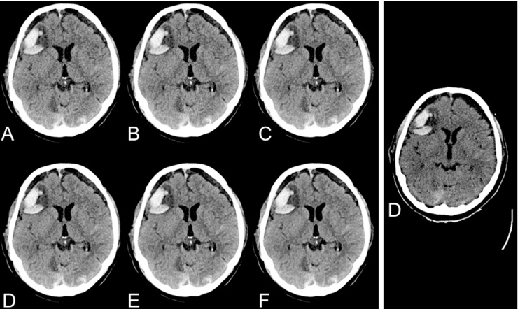
31 patients with a previous performed tilted non-contrast enhanced sequential cCT aquisition on a 4-slice CT system with only FBP reconstruction and no ATCM were prospectively enrolled in this study for a clinical indicated cCT scan. All spiral cCT examinations were performed on a 3rd generation dual-source CT system using ATCM in z-axis direction. Images were reconstructed using both, FBP and IR (level 1-5). A Monte-Carlo-simulation-based analysis was used to compare organ-specific-radiation dose. Subjective image quality for various anatomic structures was evaluated using a 4-point Likert-scale and objective image quality was evaluated by comparing signal-to-noise ratios (SNR).
Spiral cCT led to a significantly lower (p < 0.05) organ-specific-radiation dose in all targets including eye lense. Subjective image quality of spiral cCT datasets with an IR reconstruction level 5 was rated significantly higher compared to the sequential cCT acquisitions (p < 0.0001). Consecutive mean SNR was significantly higher in all spiral datasets (FBP, IR 1-5) when compared to sequential cCT with a mean SNR improvement of 44.77% (p < 0.0001).
Spiral cCT combined with ATCM and IR allows for significant-radiation dose reduction including a reduce eye lens organ-dose when compared to a tilted sequential cCT while improving subjective and objective image quality.
前瞻性评估螺旋头颅CT(cCT)联合自动管电流调制(ATCM)和迭代图像重建(IR)的图像质量和器官特异性辐射剂量,并与采用滤波反投影(FBP)重建且无ATCM的序列倾斜cCT进行比较。
本研究前瞻性纳入31例曾在4层CT系统上仅采用FBP重建且无ATCM进行过倾斜非增强序列cCT扫描的患者,进行临床指征性cCT扫描。所有螺旋cCT检查均在第三代双源CT系统上沿z轴方向使用ATCM进行。图像采用FBP和IR(1 - 5级)两种方法重建。采用基于蒙特卡洛模拟的分析方法比较器官特异性辐射剂量。使用4点李克特量表评估各种解剖结构的主观图像质量,并通过比较信噪比(SNR)评估客观图像质量。
螺旋cCT在包括晶状体在内的所有目标器官中导致显著更低(p < 0.05)的器官特异性辐射剂量。与序列cCT采集相比,IR重建5级的螺旋cCT数据集的主观图像质量评分显著更高(p < 0.0001)。与序列cCT相比,所有螺旋数据集(FBP、IR 1 - 5)的连续平均SNR均显著更高,平均SNR提高了44.77%(p < 0.0001)。
与倾斜序列cCT相比,螺旋cCT联合ATCM和IR可显著降低辐射剂量,包括降低晶状体器官剂量,同时提高主观和客观图像质量。