Fracasso Alessio, van Veluw Susanne J, Visser Fredy, Luijten Peter R, Spliet Wim, Zwanenburg Jaco J M, Dumoulin Serge O, Petridou Natalia
Experimental Psychology, Helmholtz institute, Utrecht University, Utrecht, Netherlands; Radiology, Imaging Division, University Medical Center, Utrecht, Netherlands; Spinoza Centre for Neuroimaging, Amsterdam, Netherlands.
Neurology, Brain Center Rudolf Magnus, University Medical Center, Utrecht, Netherlands.
Data Brief. 2016 Jul 5;8:990-1003. doi: 10.1016/j.dib.2016.06.058. eCollection 2016 Sep.
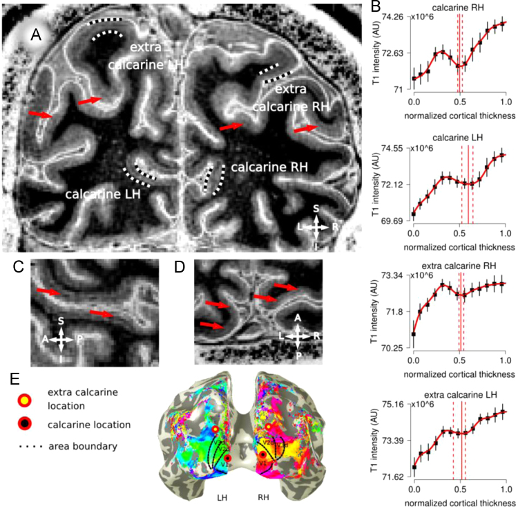
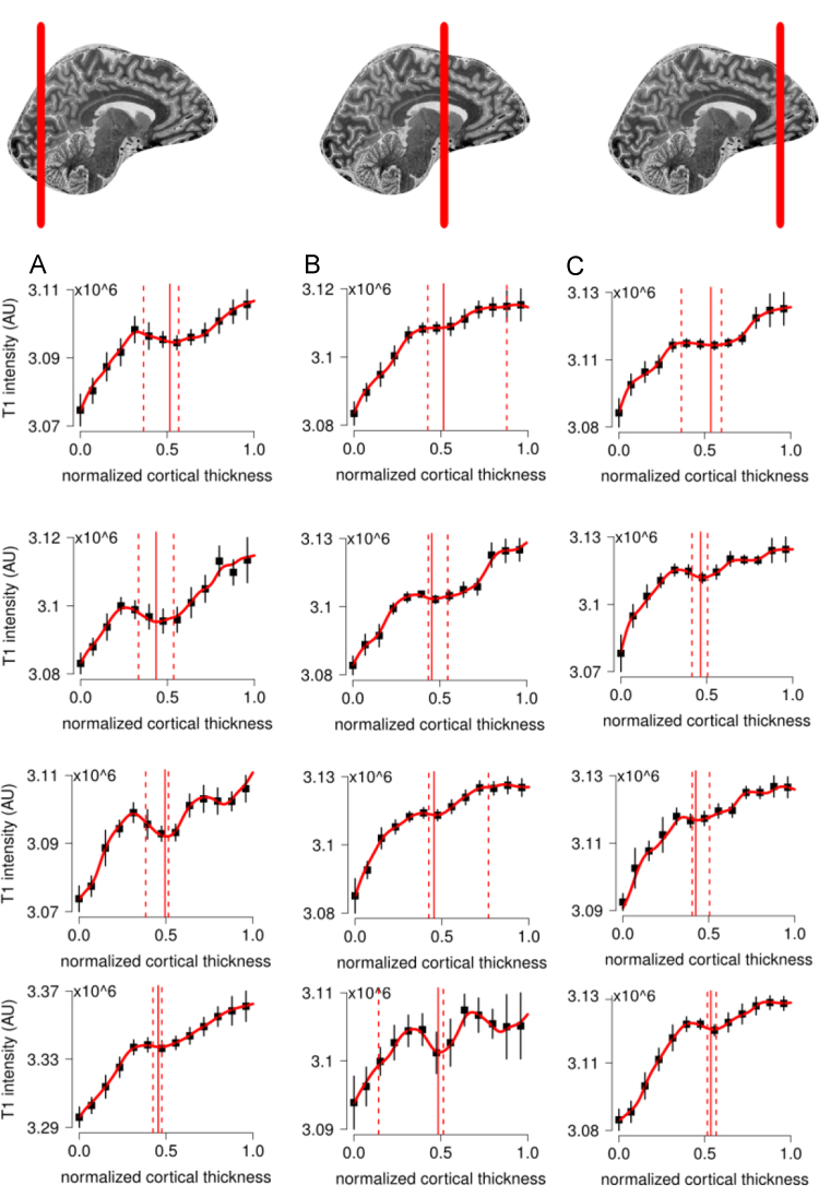
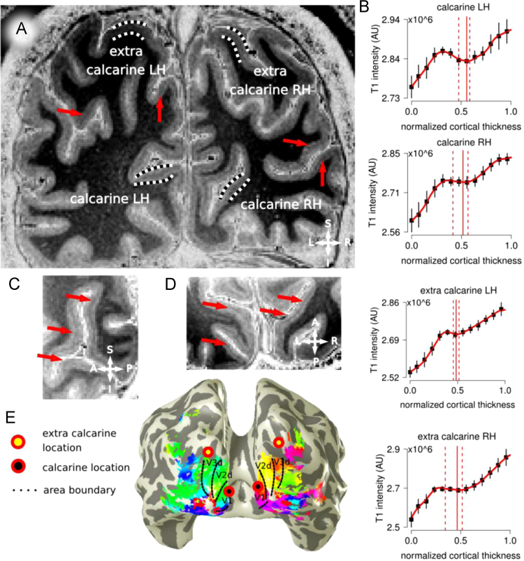
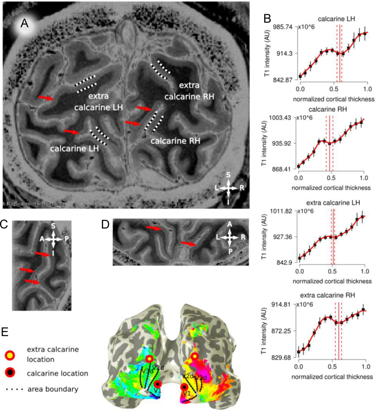
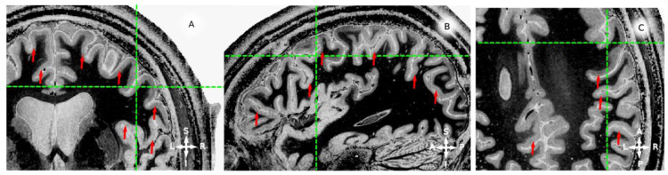
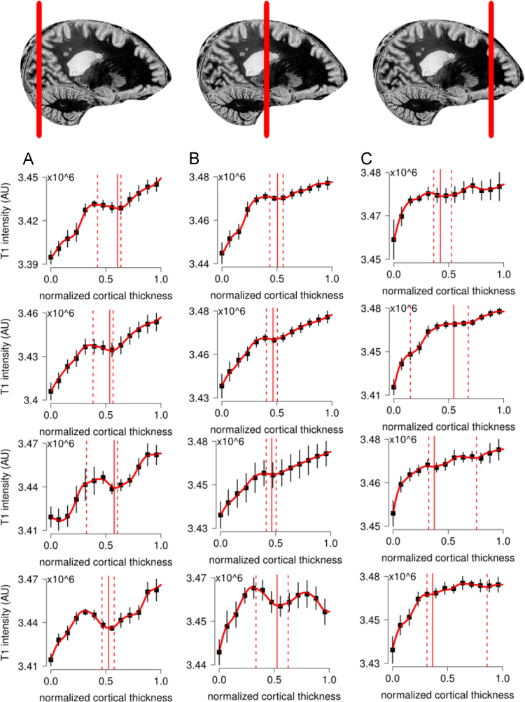
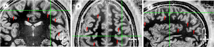
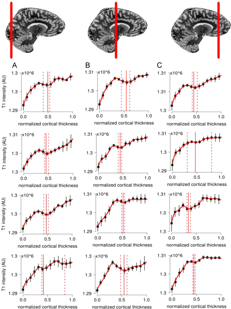
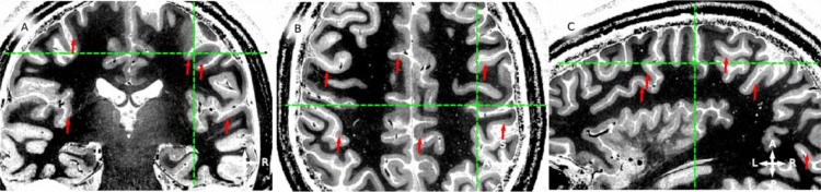
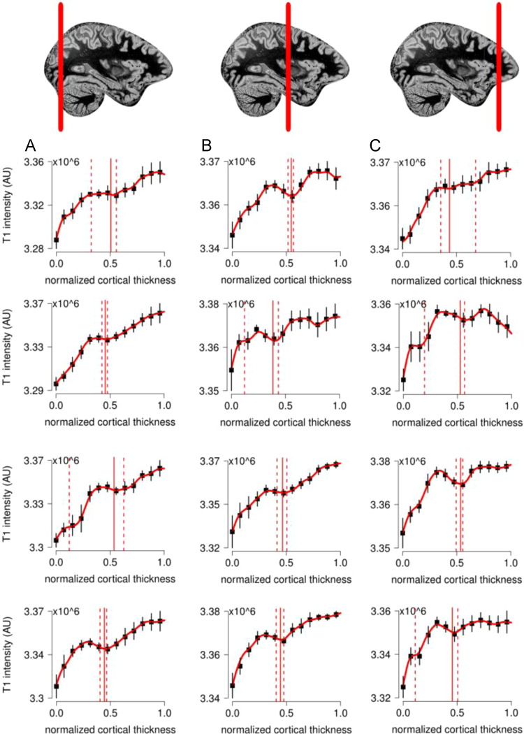
In this article we report the complete data obtained in-vivo for the paper: "Lines of Baillarger in vivo and ex-vivo: myelin contrast across lamina at 7T MRI and histology" (Fracasso et al., 2015) [1]. Single participant data (4 participants) from the occipital lobe acquisition are reported for axial, coronal and sagittal slices; early visual area functional localization and laminar profiles are reported. Data from whole brain images are reported and described (5 participants), for axial, coronal and sagittal slices. Laminar profiles from occipital, parietal and frontal lobes are reported. The data reported in this manuscript complements the paper (Fracasso et al., 2015) [1] by providing the full set of results from the complete pool of participants, on a single-participant basis. Moreover, we provide histological images from the ex-vivo sample reported in Fracasso et al. (2015) [1].
在本文中,我们报告了为论文《活体和离体状态下的贝亚尔热线条:7T磁共振成像及组织学中跨层的髓磷脂对比度》(弗拉卡索等人,2015年)[1]所获取的完整活体数据。报告了来自枕叶采集的单参与者数据(4名参与者)的轴向、冠状和矢状切片;报告了早期视觉区域功能定位和分层轮廓。报告并描述了来自全脑图像的数据(5名参与者)的轴向、冠状和矢状切片。报告了枕叶、顶叶和额叶的分层轮廓。本手稿中报告的数据通过在单参与者基础上提供来自完整参与者群体的全套结果,对论文(弗拉卡索等人,2015年)[1]进行了补充。此外,我们提供了弗拉卡索等人(2015年)[1]中报告的离体样本的组织学图像。