Yan Jianhua, Schaefferkoette Josh, Conti Maurizio, Townsend David
Department of Nuclear Medicine, First Hospital of Shanxi Medical University, 85 Jiefang S Rd, Yingze, Taiyuan, Shanxi, 030001, China.
Molecular Imaging Precision Medicine Collaborative Innovation Center, Shanxi Medical University, 85 Jiefang S Rd, Yingze, Taiyuan, Shanxi, 030001, China.
Cancer Imaging. 2016 Aug 26;16(1):26. doi: 10.1186/s40644-016-0086-0.
Lowering injected dose will have an effect on PET image quality. In this article, we aim to investigate this effect in terms of signal-to-noise ratio (SNR) in the liver, contrast-to-noise ratio (CNR) in the lesion, bias and ensemble image noise.
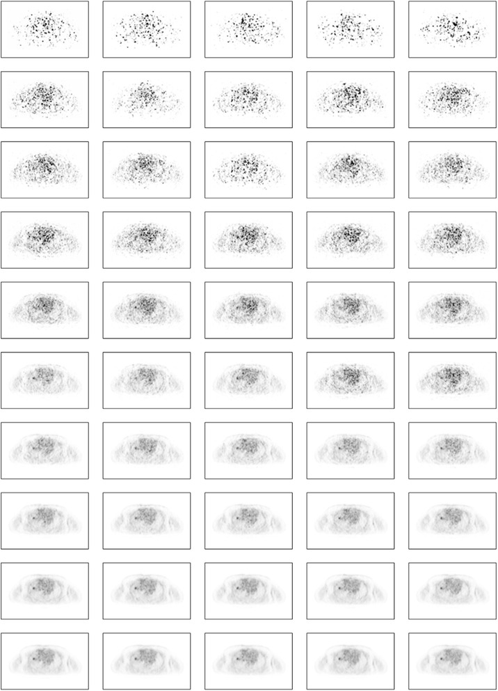
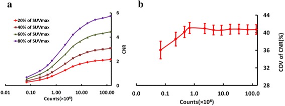
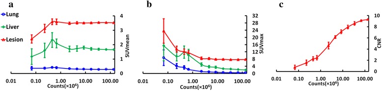
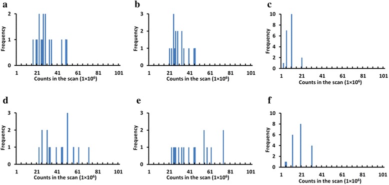
We present here our method and preliminary results using tuberculosis (TB) cases. Sixteen patients who underwent (18)F-FDG PET/MR scans covering the whole lung and portion of the liver were selected for the study. Reduced doses were simulated by randomly discarding events in the PET list mode data stream, and ten realizations at each simulated dose were generated and reconstructed. The volumes of interest (VOI) were delineated on the image reconstructed from the original full statistics data for each patient. Four thresholds (20, 40, 60 and 80 % of SUVmax) were used to quantify the effect of the threshold on CNR at the different count level. Image metrics were calculated for each VOI. This experiment allowed us to quantify the loss of SNR and CNR as a function of the counts in the scan, in turn related to dose injected. Reproducibility of mean and maximum standardized uptake value (SUVmean and SUVmax) measurement in the lesions was studied as standard deviation across 10 realizations.
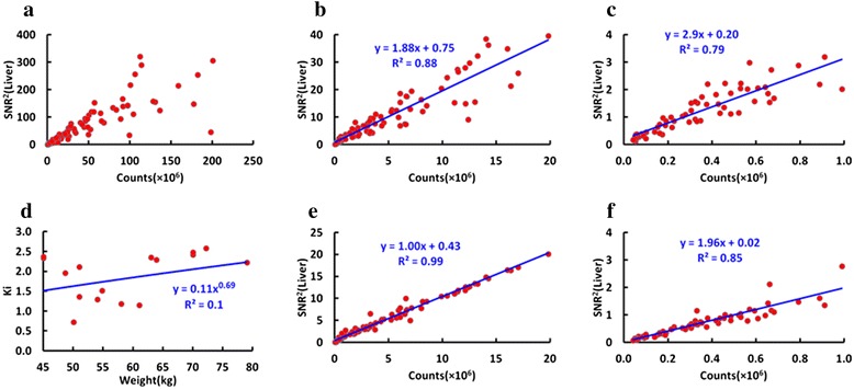
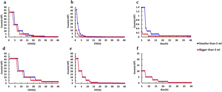
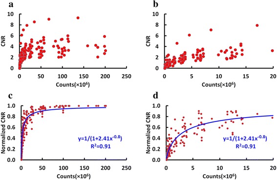
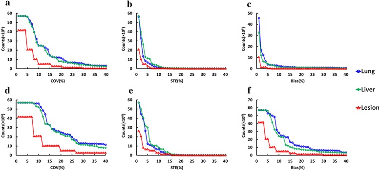
At 5 × 10(6) counts in the scan, the average SNR in the liver in the observed samples is about 3, and the CNR is reduced to 60 % of the full statistics value. The CNR in the lesion and SNR in the liver decreased with reducing count data. The variation of CNR across the four thresholds does not significantly change until the count level of 5 × 10(6). After correcting the factor related to subject's weight, the square of the SNR in the liver was found to have a very good linear relationship with detected counts. Some quantitative bias appears with count reduction. At the count level of 5 × 10(6), bias and noise in terms of SUVmean and SUVmax are up to 10 and 20 %, respectively. To keep both bias and noise less than 10 %, 5 × 10(6) counts and 20 × 10(6) counts were required for SUVmean and SUVmax, respectively.
Initial results with the given data of 16 patients diagnosed as TB demonstrated that 5 × 10(6) counts in the scan could be sufficient to yield good images in terms of SNR, CNR, bias and noise. In the future, more work needs to be done to validate the proposed method with a larger population and lung cancer patient data.
降低注射剂量会对PET图像质量产生影响。在本文中,我们旨在从肝脏的信噪比(SNR)、病变的对比噪声比(CNR)、偏差和总体图像噪声方面研究这种影响。
我们在此展示使用肺结核(TB)病例的方法和初步结果。选择16例接受了覆盖全肺和部分肝脏的(18)F-FDG PET/MR扫描的患者进行研究。通过在PET列表模式数据流中随机丢弃事件来模拟降低剂量,并在每个模拟剂量下生成并重建10次实现。在从每个患者的原始全统计数据重建的图像上勾勒出感兴趣体积(VOI)。使用四个阈值(SUVmax的20%、40%、60%和80%)来量化阈值在不同计数水平下对CNR的影响。为每个VOI计算图像指标。该实验使我们能够量化SNR和CNR的损失作为扫描中计数的函数,而计数又与注射剂量相关。研究病变中平均和最大标准化摄取值(SUVmean和SUVmax)测量的重复性作为10次实现的标准差。
在扫描中计数为5×10⁶时,观察样本中肝脏的平均SNR约为3,CNR降至全统计值的60%。随着计数数据减少,病变中的CNR和肝脏中的SNR降低。在计数水平达到5×10⁶之前,四个阈值之间CNR的变化没有显著改变。在校正与受试者体重相关的因素后,发现肝脏中SNR的平方与检测到的计数具有非常好的线性关系。随着计数减少出现一些定量偏差。在计数水平为5×10⁶时,SUVmean和SUVmax方面的偏差和噪声分别高达10%和20%。为使偏差和噪声均小于10%,SUVmean和SUVmax分别需要5×10⁶计数和20×10⁶计数。
对16例诊断为TB的患者的给定数据的初步结果表明,扫描中5×10⁶计数在SNR、CNR、偏差和噪声方面可能足以产生良好的图像。未来,需要做更多工作以用更大的人群和肺癌患者数据验证所提出的方法。