• 文献检索
  • 文档翻译
  • 深度研究
  • 学术资讯
  • Suppr Zotero 插件Zotero 插件
  • 邀请有礼
  • 套餐&价格
  • 历史记录
应用&插件
Suppr Zotero 插件Zotero 插件浏览器插件Mac 客户端Windows 客户端微信小程序
定价
高级版会员购买积分包购买API积分包
服务
文献检索文档翻译深度研究API 文档MCP 服务
关于我们
关于 Suppr公司介绍联系我们用户协议隐私条款
关注我们

Suppr 超能文献

核心技术专利:CN118964589B侵权必究
粤ICP备2023148730 号-1Suppr @ 2026

文献检索

告别复杂PubMed语法,用中文像聊天一样搜索,搜遍4000万医学文献。AI智能推荐,让科研检索更轻松。

立即免费搜索

文件翻译

保留排版,准确专业,支持PDF/Word/PPT等文件格式,支持 12+语言互译。

免费翻译文档

深度研究

AI帮你快速写综述,25分钟生成高质量综述,智能提取关键信息,辅助科研写作。

立即免费体验

用于建模目的的褐腐菌后黄丝盘菌菌丝体生长的形态学表征与定量分析

Morphological Characterization and Quantification of the Mycelial Growth of the Brown-Rot Fungus Postia placenta for Modeling Purposes.

作者信息

Du Huan, Lv Pin, Ayouz Mehdi, Besserer Arnaud, Perré Patrick

机构信息

LGPM, CentraleSupelec, Université Paris-Saclay, 92290, Châtenay-malabry, France.

ENSTIB/LERMAB, University of Lorraine, 88000, Epinal, France.

出版信息

PLoS One. 2016 Sep 7;11(9):e0162469. doi: 10.1371/journal.pone.0162469. eCollection 2016.

DOI:10.1371/journal.pone.0162469
PMID:27602575
原文链接:https://pmc.ncbi.nlm.nih.gov/articles/PMC5014427/
Abstract

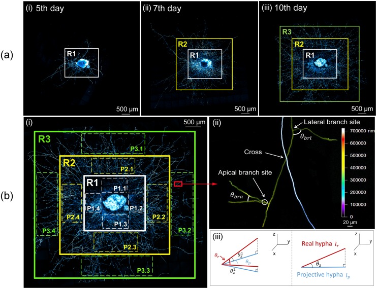
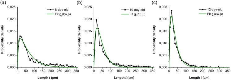
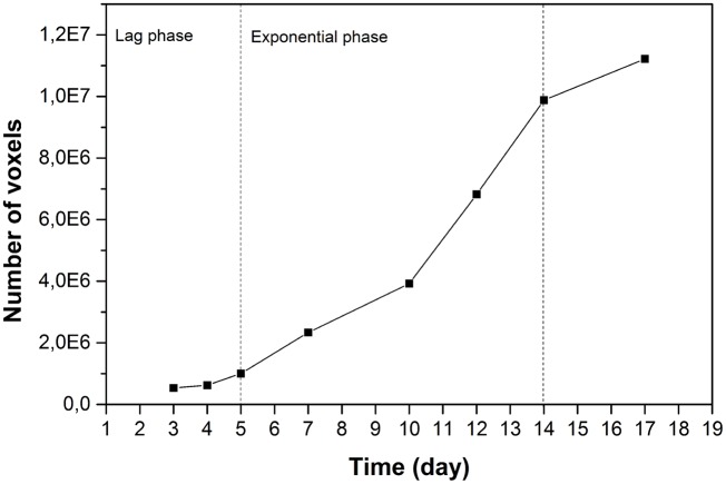
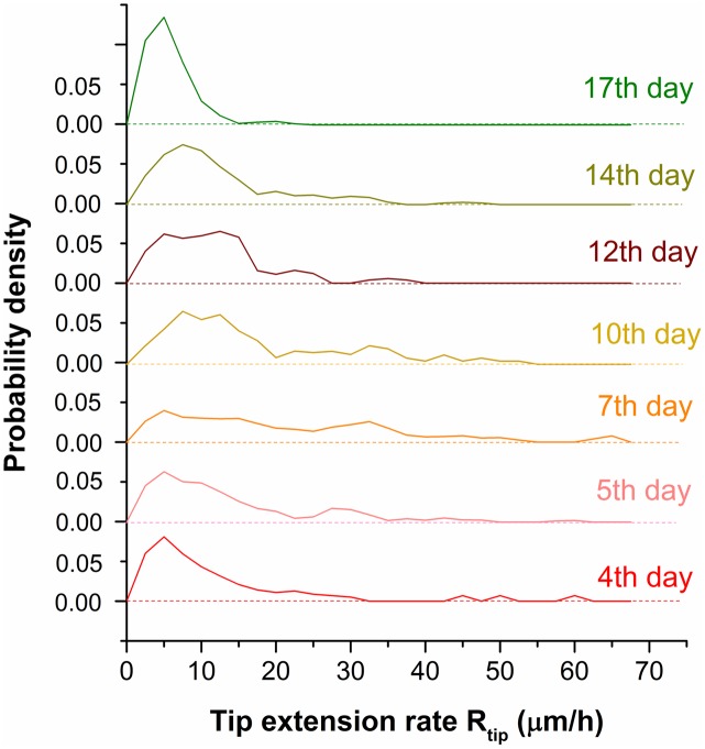
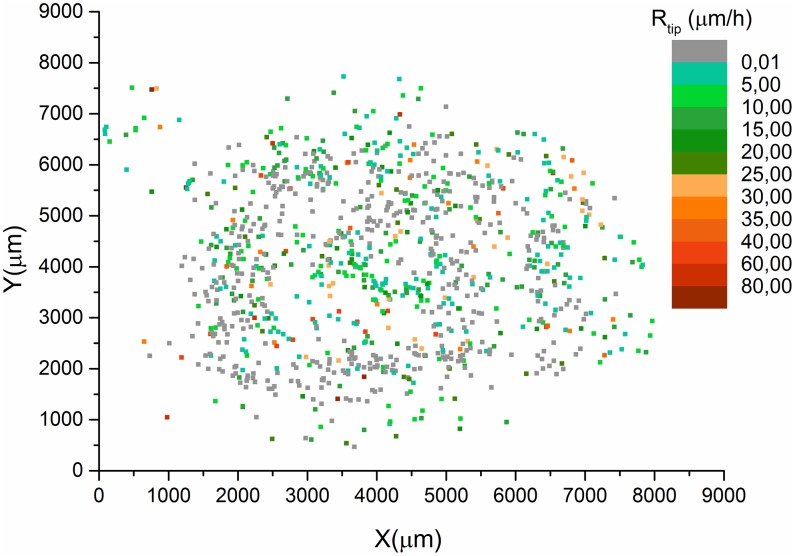
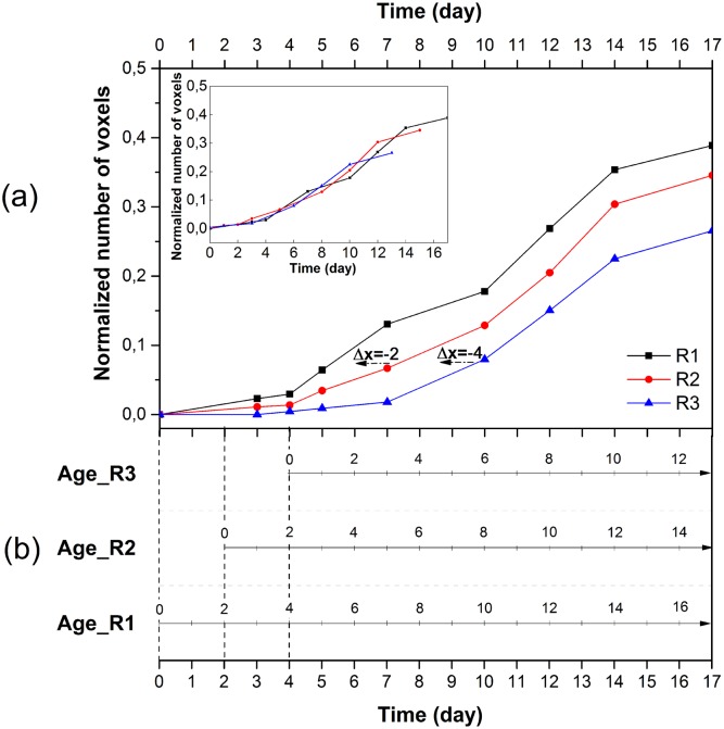
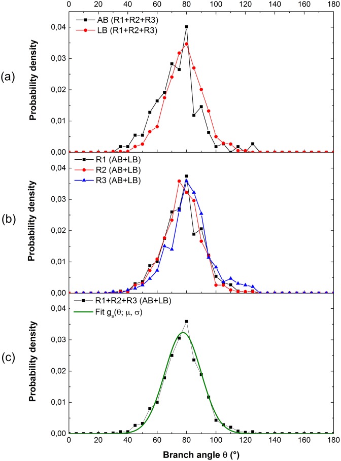
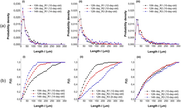
Continuous observation was performed using confocal laser scanning microscopy to visualize the three-dimensional microscopic growth of the brown-rot fungus, Postia placenta, for seventeen days. The morphological characterization of Postia placenta was quantitatively determined, including the tip extension rate, branch angle and branching length, (hyphal length between two adjacent branch sites). A voxel method has been developed to measure the growth of the biomass. Additionally, the tip extension rate distribution, the branch angle distribution and the branching length distribution, which quantified the hyphal growth characteristics, were evaluated. Statistical analysis revealed that the extension rate of tips was randomly distributed in space. The branch angle distribution did not change with the development of the colony, however, the branching length distribution did vary with the development of the colony. The experimental data will be incorporated into a lattice-based model simulating the growth of Postia placenta.

摘要

使用共聚焦激光扫描显微镜进行连续观察,以可视化褐腐菌——展齿革菌(Postia placenta)的三维微观生长,持续了17天。对展齿革菌的形态特征进行了定量测定,包括尖端延伸速率、分支角度和分支长度(两个相邻分支位点之间的菌丝长度)。已开发出一种体素方法来测量生物量的生长。此外,还评估了量化菌丝生长特征的尖端延伸速率分布、分支角度分布和分支长度分布。统计分析表明,尖端的延伸速率在空间中呈随机分布。分支角度分布不会随着菌落的发育而变化,然而,分支长度分布确实会随着菌落的发育而变化。实验数据将被纳入一个基于晶格的模型,以模拟展齿革菌的生长。

https://cdn.ncbi.nlm.nih.gov/pmc/blobs/ecb8/5014427/29cf5eeee97b/pone.0162469.g008.jpg
https://cdn.ncbi.nlm.nih.gov/pmc/blobs/ecb8/5014427/7ecd7088972d/pone.0162469.g001.jpg
https://cdn.ncbi.nlm.nih.gov/pmc/blobs/ecb8/5014427/a636ed2a9e8f/pone.0162469.g002.jpg
https://cdn.ncbi.nlm.nih.gov/pmc/blobs/ecb8/5014427/e8fef52cbe66/pone.0162469.g003.jpg
https://cdn.ncbi.nlm.nih.gov/pmc/blobs/ecb8/5014427/632f707d2968/pone.0162469.g004.jpg
https://cdn.ncbi.nlm.nih.gov/pmc/blobs/ecb8/5014427/8ba70ee79650/pone.0162469.g005.jpg
https://cdn.ncbi.nlm.nih.gov/pmc/blobs/ecb8/5014427/c98e5ddbdda4/pone.0162469.g006.jpg
https://cdn.ncbi.nlm.nih.gov/pmc/blobs/ecb8/5014427/e148d46ea5c4/pone.0162469.g007.jpg
https://cdn.ncbi.nlm.nih.gov/pmc/blobs/ecb8/5014427/29cf5eeee97b/pone.0162469.g008.jpg

相似文献

1
Morphological Characterization and Quantification of the Mycelial Growth of the Brown-Rot Fungus Postia placenta for Modeling Purposes.用于建模目的的褐腐菌后黄丝盘菌菌丝体生长的形态学表征与定量分析
PLoS One. 2016 Sep 7;11(9):e0162469. doi: 10.1371/journal.pone.0162469. eCollection 2016.
2
Hyphal morphology modification in thermal adaptation by the white-rot fungus Fomes sp. EUM1.白腐菌 Fomes sp. EUM1 通过菌丝形态改变适应热胁迫。
J Basic Microbiol. 2012 Apr;52(2):167-74. doi: 10.1002/jobm.201000528. Epub 2011 Sep 23.
3
Correction: Morphological Characterization and Quantification of the Mycelial Growth of the Brown-Rot Fungus Postia placenta for Modeling Purposes.更正:用于建模目的的褐腐菌胎盘多腔菌菌丝体生长的形态学表征与定量分析
PLoS One. 2018 May 30;13(5):e0189548. doi: 10.1371/journal.pone.0189548. eCollection 2018.
4
Localizing gene regulation reveals a staggered wood decay mechanism for the brown rot fungus Postia placenta.基因调控定位揭示了褐腐菌后垣盘革菌的交错木材腐朽机制。
Proc Natl Acad Sci U S A. 2016 Sep 27;113(39):10968-73. doi: 10.1073/pnas.1608454113. Epub 2016 Sep 12.
5
Using a grass substrate to compare decay among two clades of brown rot fungi.用草本基质比较两种褐腐真菌类群的腐朽情况。
Appl Microbiol Biotechnol. 2013 Oct;97(19):8831-40. doi: 10.1007/s00253-013-5142-0. Epub 2013 Aug 6.
6
A 3-variable PDE model for predicting fungal growth derived from microscopic mechanisms.基于微观机制预测真菌生长的 3 变量偏微分方程模型。
J Theor Biol. 2019 Jun 7;470:90-100. doi: 10.1016/j.jtbi.2019.03.015. Epub 2019 Mar 21.
7
Modelling the hyphal growth of the wood-decay fungus Physisporinus vitreus.建模木材腐朽真菌 Physisporinus vitreus 的菌丝生长。
Fungal Biol. 2011 Sep;115(9):919-32. doi: 10.1016/j.funbio.2011.06.017. Epub 2011 Jul 23.
8
Environmental scanning electron microscopic observation of the hyphal sheath and mycofibrils in Postia placenta.
Can J Microbiol. 1995 Apr-May;41(4-5):433-7. doi: 10.1139/m95-058.
9
Transcriptional profiles of laccase genes in the brown rot fungus Postia placenta MAD-R-698.褐腐菌后垣孔菌MAD-R-698中漆酶基因的转录谱
J Microbiol. 2015 Sep;53(9):606-15. doi: 10.1007/s12275-015-4705-4. Epub 2015 Aug 1.
10
Analysis of morphological relationship between micro- and macromorphology of Mortierella species using a flow-through chamber coupled with image analysis.使用流通室结合图像分析对被孢霉属物种的微观和宏观形态之间的形态关系进行分析。
J Eukaryot Microbiol. 2006 May-Jun;53(3):199-203. doi: 10.1111/j.1550-7408.2006.00094.x.

引用本文的文献

1
Full identification of a growing and branching network's spatio-temporal structures.对一个不断生长和分支的网络的时空结构进行全面识别。
Biophys J. 2025 Jan 21;124(2):284-296. doi: 10.1016/j.bpj.2024.12.002. Epub 2024 Dec 5.
2
Characterization of spatio-temporal dynamics of the constrained network of the filamentous fungus Podospora anserina using a geomatics-based approach.使用基于大地测量学的方法对丝状真菌 Podospora anserina 的约束网络的时空动态进行特征描述。
PLoS One. 2024 Feb 6;19(2):e0297816. doi: 10.1371/journal.pone.0297816. eCollection 2024.
3
Network traits predict ecological strategies in fungi.

本文引用的文献

1
Automated image-based analysis of spatio-temporal fungal dynamics.基于图像的时空真菌动态自动化分析。
Fungal Genet Biol. 2015 Nov;84:12-25. doi: 10.1016/j.fgb.2015.09.004. Epub 2015 Sep 11.
2
Modelling the hyphal growth of the wood-decay fungus Physisporinus vitreus.建模木材腐朽真菌 Physisporinus vitreus 的菌丝生长。
Fungal Biol. 2011 Sep;115(9):919-32. doi: 10.1016/j.funbio.2011.06.017. Epub 2011 Jul 23.
3
Multidimensional NMR analysis reveals truncated lignin structures in wood decayed by the brown rot basidiomycete Postia placenta.
网络特征可预测真菌的生态策略。
ISME Commun. 2022 Jan 5;2(1):2. doi: 10.1038/s43705-021-00085-1.
4
Dynamic full-field optical coherence tomography for live-cell imaging and growth-phase monitoring in .动态全场光学相干断层扫描用于. 活细胞成像和生长阶段监测。
Front Cell Infect Microbiol. 2023 Jul 12;13:1183340. doi: 10.3389/fcimb.2023.1183340. eCollection 2023.
5
Prediction and experimental evidence of different growth phases of the Podospora anserina hyphal network.预测与实验证据表明,离蠕孢霉菌丝网络存在不同的生长阶段。
Sci Rep. 2023 May 25;13(1):8501. doi: 10.1038/s41598-023-35327-w.
6
Prediction and experimental evidence of the optimisation of the angular branching process in the thallus growth of Podospora anserina.蛹草拟青霉梗枝生长中角分支过程优化的预测和实验证据。
Sci Rep. 2022 Jul 19;12(1):12351. doi: 10.1038/s41598-022-16245-9.
7
Varied solutions to multicellularity: The biophysical and evolutionary consequences of diverse intercellular bonds.多细胞性的多样解决方案:不同细胞间连接的生物物理和进化后果。
Biophys Rev (Melville). 2022 Jun;3(2):021305. doi: 10.1063/5.0080845. Epub 2022 Jun 1.
8
Morphological growth pattern of Phanerochaete chrysosporium cultivated on different Miscanthus x giganteus biomass fractions.不同芒属生物质组分上培养的黄孢原毛平革菌的形态生长模式。
BMC Microbiol. 2021 Nov 17;21(1):318. doi: 10.1186/s12866-021-02350-8.
9
Correction: Morphological Characterization and Quantification of the Mycelial Growth of the Brown-Rot Fungus Postia placenta for Modeling Purposes.更正:用于建模目的的褐腐菌胎盘多腔菌菌丝体生长的形态学表征与定量分析
PLoS One. 2018 May 30;13(5):e0189548. doi: 10.1371/journal.pone.0189548. eCollection 2018.
10
The Mycelium as a Network.菌丝体作为网络。
Microbiol Spectr. 2017 May;5(3). doi: 10.1128/microbiolspec.FUNK-0033-2017.
多维 NMR 分析揭示了由棕色腐朽担子菌胎盘 Postia 腐朽的木材中截短的木质素结构。
Environ Microbiol. 2011 Apr;13(4):1091-100. doi: 10.1111/j.1462-2920.2010.02417.x. Epub 2011 Jan 24.
4
Growth-induced mass flows in fungal networks.真菌网络中生长诱导的物质流动。
Proc Biol Sci. 2010 Nov 7;277(1698):3265-74. doi: 10.1098/rspb.2010.0735. Epub 2010 Jun 10.
5
Structural analysis of biofilms and pellets of Aspergillus niger by confocal laser scanning microscopy and cryo scanning electron microscopy.通过共聚焦激光扫描显微镜和冷冻扫描电子显微镜分析黑曲霉的生物膜和菌球的结构。
Bioresour Technol. 2010 Mar;101(6):1920-6. doi: 10.1016/j.biortech.2009.10.036.
6
Solubilization of calcium phosphate as a consequence of carbon translocation by Rhizoctonia solani.由于 Rhizoctonia solani 的碳转移,导致磷酸钙溶解。
FEMS Microbiol Ecol. 2002 Apr 1;40(1):65-71. doi: 10.1111/j.1574-6941.2002.tb00937.x.
7
Fine structure and X-ray microanalysis of silicified woods from a Tertiary basin Pohang, Korea by scanning electron microscopy.
Micron. 2009 Jul-Aug;40(5-6):519-25. doi: 10.1016/j.micron.2009.04.006. Epub 2009 May 3.
8
Branching of fungal hyphae: regulation, mechanisms and comparison with other branching systems.真菌菌丝的分支:调控、机制及与其他分支系统的比较
Mycologia. 2008 Nov-Dec;100(6):823-32. doi: 10.3852/08-177.
9
Frozen in time: a new method using cryo-scanning electron microscopy to visualize root-fungal interactions.
New Phytol. 2006;172(2):369-74. doi: 10.1111/j.1469-8137.2006.01825.x.
10
Two new fluorescent dyes applicable for visualization of fungal cell walls.两种适用于真菌细胞壁可视化的新型荧光染料。
Mycologia. 2005 May-Jun;97(3):580-8. doi: 10.3852/mycologia.97.3.580.