Salö Martin, Marungruang Nittaya, Roth Bodil, Sundberg Tiia, Stenström Pernilla, Arnbjörnsson Einar, Fåk Frida, Ohlsson Bodil
Department of Clinical Sciences, Pediatrics, Lund University, Lund, Sweden.
Department of Pediatric Surgery, Skåne University Hospital, Lund University, Lasarettsgatan 48, 221 85, Lund, Sweden.
Int J Colorectal Dis. 2017 Jan;32(1):19-28. doi: 10.1007/s00384-016-2639-x. Epub 2016 Sep 9.
BACKGROUND/AIM: The role of the microbiome has been widely discussed in the etiology of appendicitis. The primary aim was to evaluate the microbiome in the normal appendix and in appendicitis specifically divided into the three clinically and histopathologically defined grades of inflammation. Secondary aims were to examine whether there were any microbiome differences between proximal and distal appendices, and relate the microbiome with histopathological findings.
A prospective pilot study was conducted of children undergoing appendectomy for appendicitis. The diagnosis was based on histopathological analysis. Children with incidental appendectomy were used as controls. The proximal and distal mucosa from the appendices were analyzed with 16S rRNA gene sequencing.
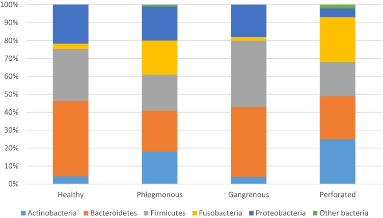
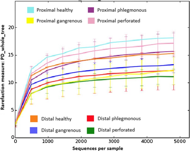
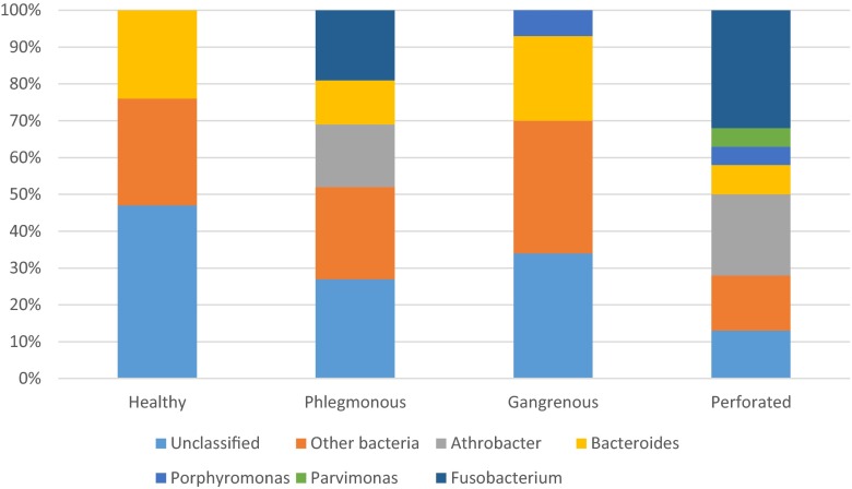
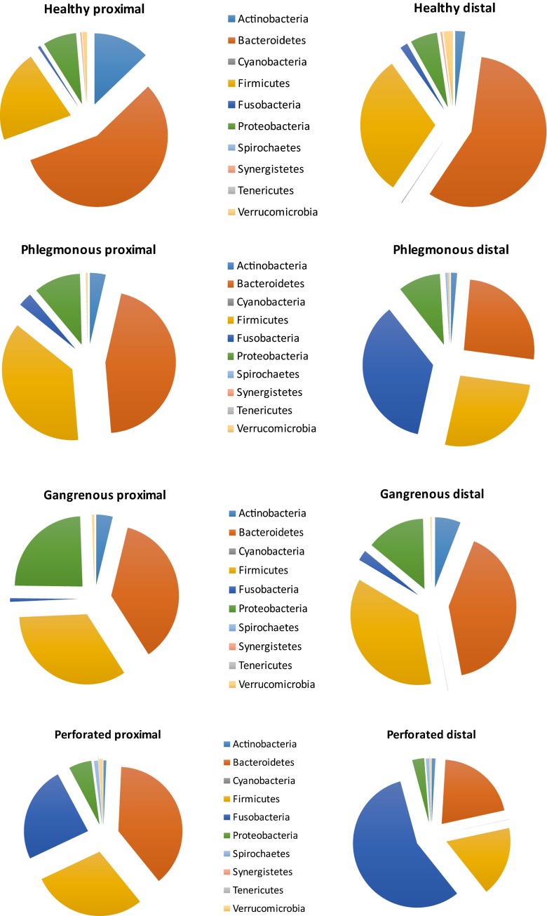
A total of 22 children, 3 controls and 19 appendicitis patients; 11 phlegmonous, 4 gangrenous, and 4 perforated appendices, were prospectively included. The amount of Fusobacterium increased and Bacteroides decreased in phlegmonous and perforated appendicitis compared to controls, but statistical significance was not reached, and this pattern was not seen in gangrenous appendicitis. No relation could be seen between different bacteria and the grade of inflammation, and there was a wide variation of abundances at phylum, genus, and species level within every specific group of patients. Further, no significant differences could be detected when comparing the microbiome in proximal and distal mucosa, which may be because the study was underpowered. A trend with more abundance of Fusobacteria in the distal mucosa was seen in appendicitis patients with obstruction (25 and 13 %, respectively, p = 0.06).
The pattern of microbiome differed not only between groups, but also within groups. However, no statistically significant differences could be found in the microbiome between groups or clinical conditions. No correlation between a specific bacteria and grade of inflammation was found. In the vast majority of cases of appendicitis, changes in microbiome do not seem to be the primary event. Since there seem to be differences in microbiome patterns depending on the sample site, the exact localization of biopsy sampling must be described in future studies.
背景/目的:微生物群在阑尾炎病因学中的作用已得到广泛讨论。主要目的是评估正常阑尾以及根据临床和组织病理学分为三个炎症等级的阑尾炎中的微生物群。次要目的是检查近端和远端阑尾之间是否存在微生物群差异,并将微生物群与组织病理学结果相关联。
对因阑尾炎接受阑尾切除术的儿童进行了一项前瞻性试点研究。诊断基于组织病理学分析。将接受偶然阑尾切除术的儿童用作对照。使用16S rRNA基因测序分析阑尾的近端和远端黏膜。
前瞻性纳入了22名儿童,其中3名对照和19名阑尾炎患者;11例蜂窝织炎性、4例坏疽性和4例穿孔性阑尾。与对照相比,蜂窝织炎性和穿孔性阑尾炎中具核梭杆菌数量增加而拟杆菌数量减少,但未达到统计学意义,且在坏疽性阑尾炎中未观察到这种模式。不同细菌与炎症等级之间未见关联,并且在每组特定患者中,门、属和种水平的丰度存在很大差异。此外,比较近端和远端黏膜中的微生物群时未检测到显著差异,这可能是因为该研究的效能不足。在有梗阻的阑尾炎患者中,远端黏膜中具核梭杆菌丰度更高的趋势较为明显(分别为25%和13%,p = 0.06)。
微生物群模式不仅在组间不同,而且在组内也不同。然而,在组间或临床情况之间的微生物群中未发现统计学上的显著差异。未发现特定细菌与炎症等级之间存在相关性。在绝大多数阑尾炎病例中,微生物群的变化似乎不是主要事件。由于根据样本部位微生物群模式似乎存在差异,未来研究必须描述活检采样的确切定位。