Aguado Carolina, García-Madrona Sebastián, Gil-Minguez Mercedes, Luján Rafael
Synaptic Structure Laboratory, Department Ciencias Médicas, Instituto de Investigación en Discapacidades Neurológicas (IDINE), Facultad de Medicina, Universidad Castilla-La Mancha Albacete, Spain.
Front Neuroanat. 2016 Aug 26;10:83. doi: 10.3389/fnana.2016.00083. eCollection 2016.
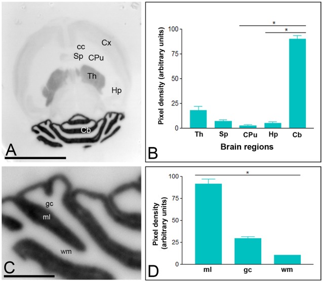
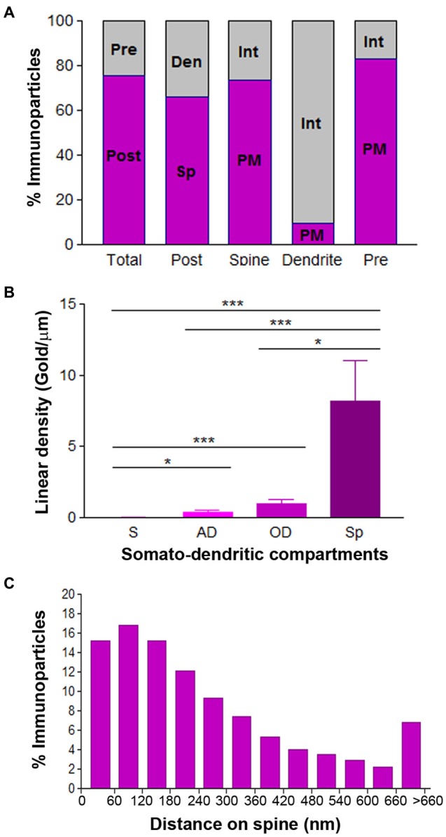
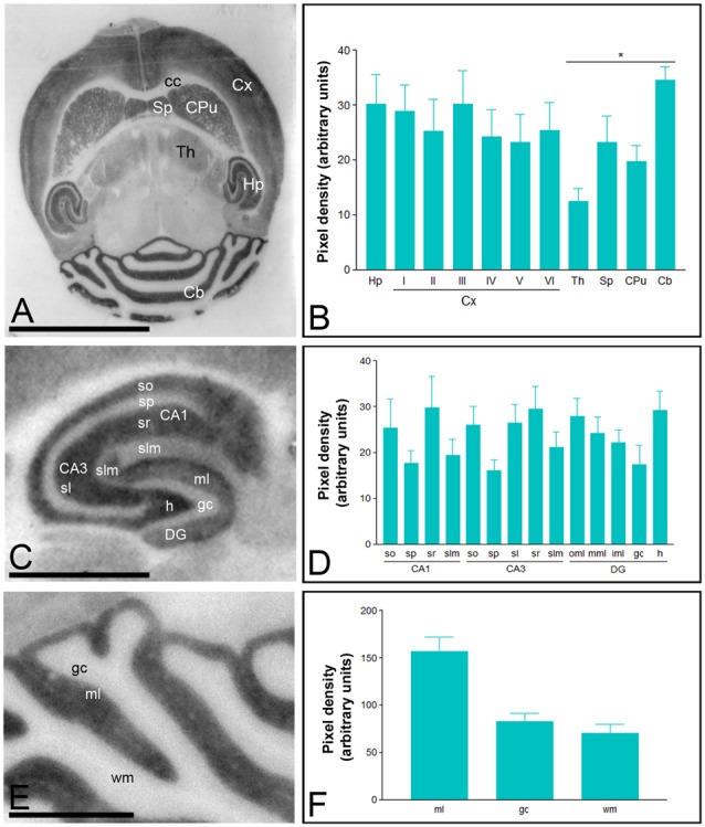
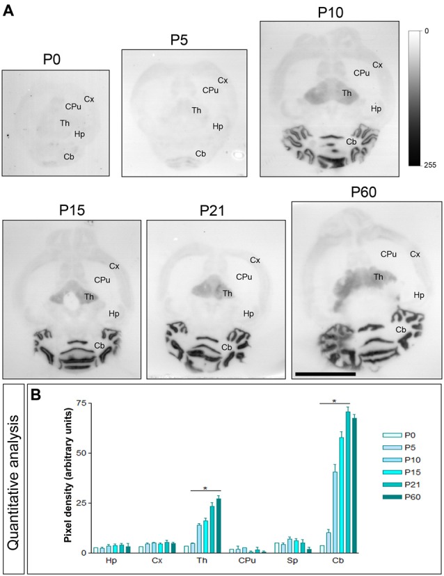
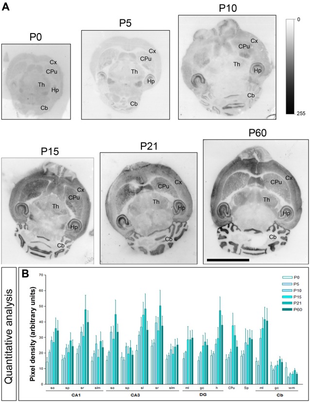
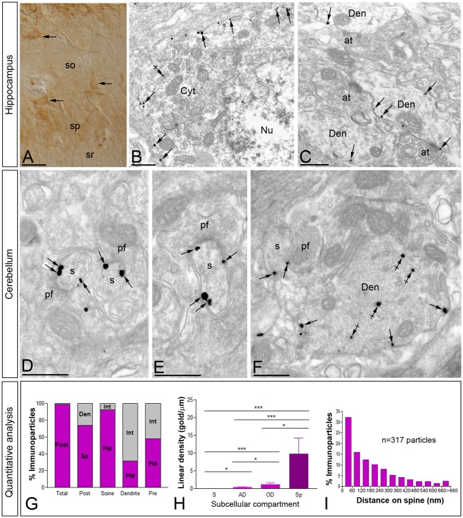
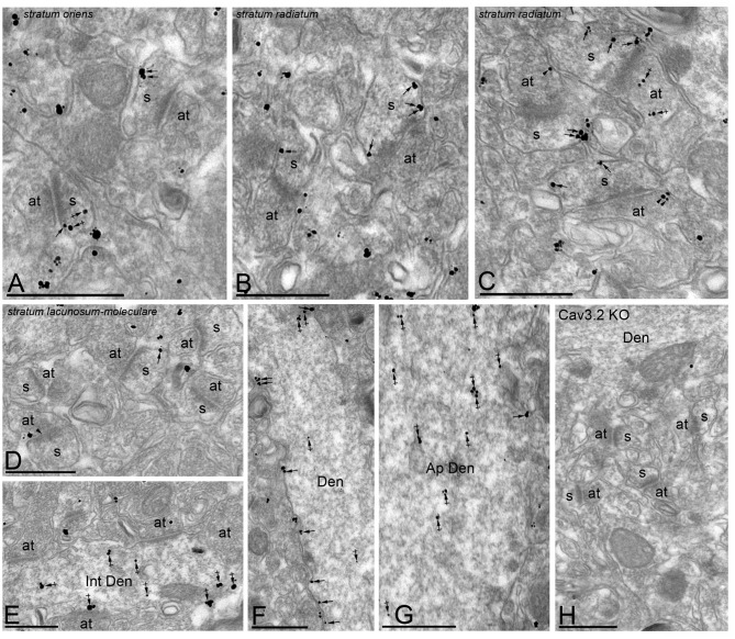
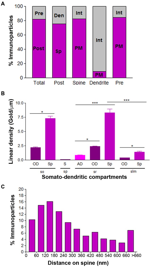
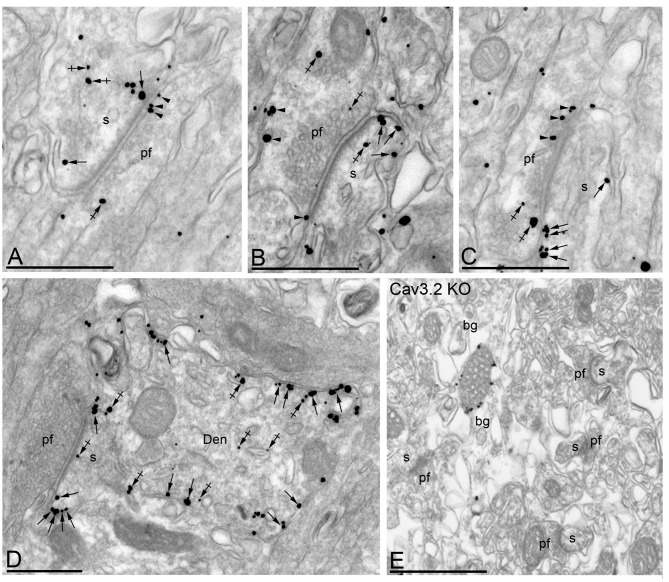
T-type calcium (Ca(2+)) channels play a central role in regulating membrane excitability in the brain. Although the contributions of T-type current to neuron output is often proposed to reflect a differential distribution of T-type channel subtypes to somato-dendritic compartments, their precise subcellular distributions in central neurons are not fully determined. Using histoblot and high-resolution immunoelectron microscopic techniques, we have investigated the expression, regional distribution and subcellular localization of T-type Cav3.1 and Cav3.2 channel subunits in the adult brain, as well as the ontogeny of expression during postnatal development. Histoblot analysis showed that Cav3.1 and Cav3.2 proteins were widely expressed in the brain, with mostly non-overlapping patterns. Cav3.1 showed the highest expression level in the molecular layer (ml) of the cerebellum (Cb), and Cav3.2 in the hippocampus (Hp) and the ml of Cb. During development, levels of Cav3.1 and Cav3.2 increased with age, although there were marked region- and developmental stage-specific differences in their expression. At the cellular and subcellular level, immunoelectron microscopy showed that labeling for Cav3.1 was present in somato-dendritic domains of hippocampal interneurons and Purkinje cells (PCs), while Cav3.2 was present in somato-dendritic domains of CA1 pyramidal cells, hippocampal interneurons and PCs. Most of the immunoparticles for Cav3.1 and Cav3.2 were either associated with the plasma membrane or the intracellular membranes, with notable differences depending on the compartment. Thus, Cav3.1 was mainly located in the plasma membrane of interneurons, whereas Cav3.2 was mainly located in the plasma membrane of dendritic spines and had a major intracellular distribution in dendritic shafts. In PCs, Cav3.1 and Cav3.2 showed similar distribution patterns. In addition to its main postsynaptic distribution, Cav3.2 but not Cav3.1 was also detected in axon terminals establishing excitatory synapses. These results shed new light on the subcellular localization of T-type channel subunits and provide evidence for the non-uniform distribution of Cav3.1 and Cav3.2 subunits over the plasma membrane of central neurons, which may account for the functional heterogeneity of T-type mediated current.
T型钙(Ca(2+))通道在调节大脑膜兴奋性方面起着核心作用。尽管通常认为T型电流对神经元输出的贡献反映了T型通道亚型在胞体-树突状区室的差异分布,但它们在中枢神经元中精确的亚细胞分布尚未完全确定。我们使用组织印迹和高分辨率免疫电子显微镜技术,研究了T型Cav3.1和Cav3.2通道亚基在成人大脑中的表达、区域分布和亚细胞定位,以及出生后发育过程中表达的个体发生。组织印迹分析表明,Cav3.1和Cav3.2蛋白在大脑中广泛表达,且大多呈非重叠模式。Cav3.1在小脑(Cb)分子层(ml)中的表达水平最高,而Cav3.2在海马体(Hp)和Cb的ml中表达最高。在发育过程中,Cav3.1和Cav3.2的水平随年龄增加,尽管它们的表达存在明显的区域和发育阶段特异性差异。在细胞和亚细胞水平上,免疫电子显微镜显示,海马中间神经元和浦肯野细胞(PCs)的胞体-树突状区域存在Cav3.1的标记,而Cav3.2存在于CA1锥体细胞、海马中间神经元和PCs的胞体-树突状区域。Cav3.1和Cav3.2的大多数免疫颗粒与质膜或细胞内膜相关,具体取决于区室,存在显著差异。因此,Cav3.1主要位于中间神经元的质膜中,而Cav3.2主要位于树突棘的质膜中,并且在树突轴中有主要的细胞内分布。在PCs中,Cav3.1和Cav3.2表现出相似的分布模式。除了主要的突触后分布外,在建立兴奋性突触的轴突终末也检测到了Cav3.2,而未检测到Cav3.1。这些结果为T型通道亚基的亚细胞定位提供了新的线索,并为Cav3.1和Cav3.2亚基在中枢神经元质膜上的非均匀分布提供了证据,这可能解释了T型介导电流的功能异质性。