Fernandez-Leiro Rafael, Scheres Sjors H W
MRC Laboratory of Molecular Biology, Cambridge Biomedical Campus, Cambridge CB2 0QH, UK.
Nature. 2016 Sep 15;537(7620):339-46. doi: 10.1038/nature19948.
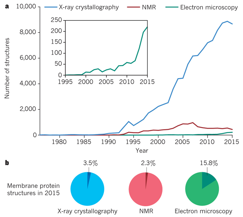
Knowledge of the three-dimensional structures of proteins and other biological macromolecules often aids understanding of how they perform complicated tasks in the cell. Because many such tasks involve the cleavage or formation of chemical bonds, structural characterization at the atomic level is most useful. Developments in the electron microscopy of frozen hydrated samples (cryo-electron microscopy) are providing unprecedented opportunities for the structural characterization of biological macromolecules. This is resulting in a wave of information about processes in the cell that were impossible to characterize with existing techniques in structural biology.
了解蛋白质和其他生物大分子的三维结构通常有助于理解它们如何在细胞中执行复杂任务。由于许多此类任务涉及化学键的断裂或形成,原子水平的结构表征最为有用。冷冻水合样品的电子显微镜技术(冷冻电子显微镜)的发展为生物大分子的结构表征提供了前所未有的机会。这正带来一波关于细胞内过程的信息,而这些过程是现有结构生物学技术无法表征的。