• 文献检索
  • 文档翻译
  • 深度研究
  • 学术资讯
  • Suppr Zotero 插件Zotero 插件
  • 邀请有礼
  • 套餐&价格
  • 历史记录
应用&插件
Suppr Zotero 插件Zotero 插件浏览器插件Mac 客户端Windows 客户端微信小程序
定价
高级版会员购买积分包购买API积分包
服务
文献检索文档翻译深度研究API 文档MCP 服务
关于我们
关于 Suppr公司介绍联系我们用户协议隐私条款
关注我们

Suppr 超能文献

核心技术专利:CN118964589B侵权必究
粤ICP备2023148730 号-1Suppr @ 2026

文献检索

告别复杂PubMed语法,用中文像聊天一样搜索,搜遍4000万医学文献。AI智能推荐,让科研检索更轻松。

立即免费搜索

文件翻译

保留排版,准确专业,支持PDF/Word/PPT等文件格式,支持 12+语言互译。

免费翻译文档

深度研究

AI帮你快速写综述,25分钟生成高质量综述,智能提取关键信息,辅助科研写作。

立即免费体验

比较口腔内放射摄影和计算机断层扫描检测犬牙周炎和牙髓病的放射影像学征象:一项一致性研究。

Comparing Intraoral Radiography and Computed Tomography for Detecting Radiographic Signs of Periodontitis and Endodontic Disease in Dogs: An Agreement Study.

机构信息

Department of Clinical Sciences, College of Veterinary Medicine, Cornell University , Ithaca, NY , USA.

出版信息

Front Vet Sci. 2016 Aug 31;3:68. doi: 10.3389/fvets.2016.00068. eCollection 2016.

DOI:10.3389/fvets.2016.00068
PMID:27630993
原文链接:https://pmc.ncbi.nlm.nih.gov/articles/PMC5005414/
Abstract

OBJECTIVE

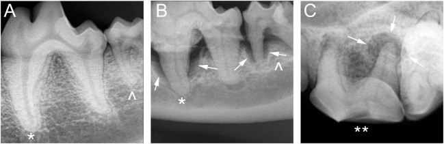
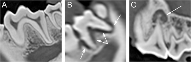
To determine whether computed tomography (CT) and intraoral radiography are interchangeable for detecting signs of periodontitis and endodontic disease in dogs.

MATERIALS AND METHODS

An agreement study was performed using 40 dogs that previously underwent intraoral radiography and CT during the same anesthetic episode. Images of each tooth were examined by two blinded observers for signs of periodontitis and/or endodontic disease. Agreement between imaging modalities and between observers was assessed using the Kappa statistic.

RESULTS

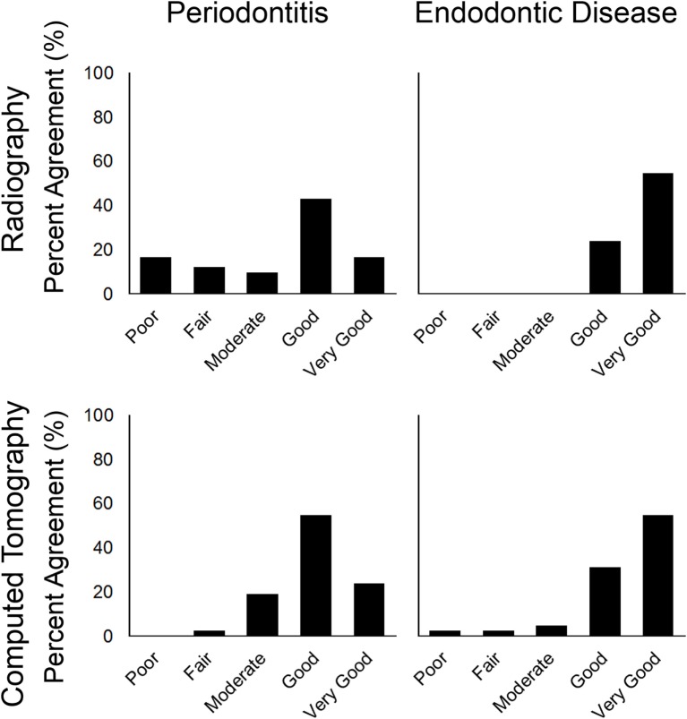
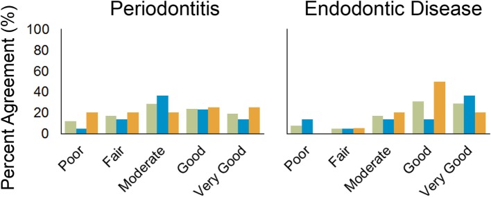
Agreement between modalities for detecting periodontitis in the maxillae ranged from poor to very good (κ 0.07-1.00) with 16/20 (80%) of the teeth having a score of moderate or better (κ ≥ 0.41). Agreement between modalities for detecting signs consistent with periodontitis in the mandibles ranged from poor to very good (κ 0.01-1.00) with 10/22 (45%) of the teeth having a score of good or better (κ ≥ 0.61); 50% of the disagreement was present in the incisors. Agreement between modalities for detecting signs consistent with endodontic disease in the whole mouth ranged from fair to very good (κ 0.21-1.00) with 30/42 (71%) of the teeth having a score of moderate or better (κ ≥ 0.41). Agreement between observers evaluating intraoral radiology ranged from poor to very good (κ 0.05-1) for detecting signs consistent with periodontitis and from fair to very good (κ 0.36-1) for detecting signs consistent with endodontic disease, in the whole mouth. Agreement between observers evaluating CT ranged from fair to very good (κ 0.35-1) for detecting signs consistent with periodontitis and from fair to very good (κ 0.36-1) for detecting signs consistent with endodontic disease, in the whole mouth.

CONCLUSION

Performing both CT and intraoral radiography may be unnecessary to detect signs consistent with periodontitis and endodontic disease in dogs based on the amount of agreement between modalities and observers when CT images are acquired and reconstructed in 0.5 or 1 mm slice thickness, except for diagnosing periodontitis in the mandibular incisors.

摘要

目的

确定计算机断层扫描(CT)和口腔内放射摄影术是否可互换用于检测犬牙周炎和牙髓疾病的迹象。

材料和方法

使用 40 只先前在同一麻醉期内接受过口腔内放射摄影术和 CT 的犬进行了一项一致性研究。两名盲法观察者检查每颗牙齿的图像,以检测牙周炎和/或牙髓疾病的迹象。使用 Kappa 统计评估两种成像方式之间以及观察者之间的一致性。

结果

上颌骨中,CT 与口腔内放射摄影术检测牙周炎的一致性范围从差到极好(κ 0.07-1.00),其中 16/20(80%)颗牙齿的评分中度或更好(κ≥0.41)。下颌骨中 CT 与口腔内放射摄影术检测与牙周炎一致的迹象的一致性范围从差到极好(κ 0.01-1.00),其中 10/22(45%)颗牙齿的评分较好或更好(κ≥0.61);50%的差异存在于切牙中。整个口腔中 CT 与口腔内放射摄影术检测与牙髓疾病一致的迹象的一致性范围从尚可到极好(κ 0.21-1.00),其中 30/42(71%)颗牙齿的评分中度或更好(κ≥0.41)。评估口腔内放射学的两名观察者之间的一致性范围为差到极好(κ 0.05-1),用于检测与牙周炎一致的迹象,以及尚可到极好(κ 0.36-1),用于检测与牙髓疾病一致的迹象,在整个口腔中。评估 CT 的两名观察者之间的一致性范围为差到极好(κ 0.35-1),用于检测与牙周炎一致的迹象,以及尚可到极好(κ 0.36-1),用于检测与牙髓疾病一致的迹象,在整个口腔中。

结论

根据两种成像方式和观察者之间的一致性程度,在获得 CT 图像并以 0.5 或 1 毫米切片厚度重建时,除了诊断下颌切牙的牙周炎外,对犬进行 CT 和口腔内放射摄影术可能都没有必要来检测与牙周炎和牙髓疾病一致的迹象。

https://cdn.ncbi.nlm.nih.gov/pmc/blobs/7438/5005414/d2462a07f886/fvets-03-00068-g004.jpg
https://cdn.ncbi.nlm.nih.gov/pmc/blobs/7438/5005414/5d57fb6c20d6/fvets-03-00068-g001.jpg
https://cdn.ncbi.nlm.nih.gov/pmc/blobs/7438/5005414/1a1a06573ea7/fvets-03-00068-g002.jpg
https://cdn.ncbi.nlm.nih.gov/pmc/blobs/7438/5005414/9a2cac5159f7/fvets-03-00068-g003.jpg
https://cdn.ncbi.nlm.nih.gov/pmc/blobs/7438/5005414/d2462a07f886/fvets-03-00068-g004.jpg

相似文献

1
Comparing Intraoral Radiography and Computed Tomography for Detecting Radiographic Signs of Periodontitis and Endodontic Disease in Dogs: An Agreement Study.比较口腔内放射摄影和计算机断层扫描检测犬牙周炎和牙髓病的放射影像学征象:一项一致性研究。
Front Vet Sci. 2016 Aug 31;3:68. doi: 10.3389/fvets.2016.00068. eCollection 2016.
2
Interpretation of chemically created periapical lesions using 2 different dental cone-beam computerized tomography units, an intraoral digital sensor, and conventional film.使用2种不同的牙科锥形束计算机断层扫描设备、口腔内数字传感器和传统胶片对化学性根尖周病变进行解读。
Oral Surg Oral Med Oral Pathol Oral Radiol Endod. 2009 Mar;107(3):426-32. doi: 10.1016/j.tripleo.2008.08.017. Epub 2008 Nov 8.
3
Comparison of intraoral radiography and cone-beam computed tomography for the detection of periodontal defects: an in vitro study.口腔内放射摄影术与锥形束计算机断层扫描术在检测牙周缺损方面的比较:一项体外研究。
BMC Oral Health. 2015 May 28;15:64. doi: 10.1186/s12903-015-0046-2.
4
Comparison of the accuracy of periapical radiography with CBCT taken at 3 different voxel sizes in detecting simulated endodontic complications: an ex vivo study.不同体素大小锥形束 CT 与传统根尖片在检测模拟根管治疗并发症准确性的比较:一项离体研究。
Dentomaxillofac Radiol. 2018 May;47(4):20170399. doi: 10.1259/dmfr.20170399. Epub 2018 Feb 22.
5
Cone-beam computerized tomographic, radiographic, and histologic evaluation of periapical repair in dogs' post-endodontic treatment.犬牙髓治疗后根尖周修复的锥形束计算机断层扫描、放射成像及组织学评估
Oral Surg Oral Med Oral Pathol Oral Radiol Endod. 2009 Nov;108(5):796-805. doi: 10.1016/j.tripleo.2009.06.016. Epub 2009 Sep 5.
6
Cone-beam Computed Tomography Compared with Intraoral Radiographic Lesions in Endodontic Outcome Studies: A Systematic Review.锥形束计算机断层扫描与口腔内放射学病变在牙髓治疗结局研究中的比较:系统评价。
J Endod. 2018 Nov;44(11):1626-1631. doi: 10.1016/j.joen.2018.08.006.
7
Application of computerised image analysis to monitoring endodontic therapy: reproducibility and comparison with visual assessment.
Dentomaxillofac Radiol. 1997 Mar;26(2):79-84. doi: 10.1038/sj.dmfr.4600207.
8
Effectiveness of limited cone-beam computed tomography in the detection of horizontal root fracture.有限锥形束计算机断层扫描在检测水平根折中的有效性
Dent Traumatol. 2009 Jun;25(3):256-61. doi: 10.1111/j.1600-9657.2009.00770.x.
9
Interobserver agreement of computed tomography in detecting calcified intervertebral discs in comparison with radiography in a population of 13 healthy British Dachshund dogs.在13只健康英国腊肠犬群体中,比较计算机断层扫描与X线摄影检测钙化椎间盘时的观察者间一致性。
Vet Rec Open. 2023 Mar 28;10(1):e59. doi: 10.1002/vro2.59. eCollection 2023 Jun.
10
Diagnostic accuracy of small volume cone beam computed tomography and intraoral periapical radiography for the detection of simulated external inflammatory root resorption.小视野锥形束 CT 与口腔根尖片诊断模拟外吸收性根面牙骨质吸收的准确性比较
Int Endod J. 2011 Feb;44(2):136-47. doi: 10.1111/j.1365-2591.2010.01819.x. Epub 2010 Nov 17.

引用本文的文献

1
Management of dental disease in eared seals (family Otariidae): a Delphi approach.海狗科动物牙病的管理:德尔菲法
Front Vet Sci. 2025 Aug 26;12:1619326. doi: 10.3389/fvets.2025.1619326. eCollection 2025.
2
Evaluation of Feline Permanent Canine Tooth Mineral Density Using Micro-Computed Tomography.使用微型计算机断层扫描评估猫恒犬齿的矿物质密度
Vet Sci. 2023 Mar 12;10(3):217. doi: 10.3390/vetsci10030217.
3
Computed tomography features of presumed normal mandibular and medial retropharyngeal lymph nodes in dogs.犬下颌和咽后内侧正常淋巴结的计算机断层扫描特征。

本文引用的文献

1
Radiologic Assessment of the Periodontal Patient.牙周病患者的放射学评估
Dent Clin North Am. 2016 Jan;60(1):91-104. doi: 10.1016/j.cden.2015.08.003.
2
Oral and Maxillofacial Imaging.口腔颌面影像学
Dent Clin North Am. 2016 Jan;60(1):1-37. doi: 10.1016/j.cden.2015.08.001. Epub 2015 Oct 21.
3
Comparison of the Diagnostic Image Quality of the Canine Maxillary Dentoalveolar Structures Obtained by Cone Beam Computed Tomography and 64-Multidetector Row Computed Tomography.锥形束计算机断层扫描和64排多层螺旋计算机断层扫描获得的犬上颌牙牙槽结构诊断图像质量的比较
Can J Vet Res. 2022 Jan;86(1):27-34.
4
Comparison of dental radiography and computed tomography: measurement of dentoalveolar structures in healthy, small-sized dogs and cats.牙科X线摄影与计算机断层扫描的比较:健康小型犬猫牙槽结构的测量
J Vet Sci. 2020 Sep;21(5):e75. doi: 10.4142/jvs.2020.21.e75.
J Vet Dent. 2015 Summer;32(2):80-6. doi: 10.1177/089875641503200201.
4
Cone beam computed tomography - know its secrets.锥形束计算机断层扫描——了解其奥秘。
J Int Oral Health. 2015 Feb;7(2):64-8.
5
Review article: Maxillofacial emergencies: oral pain and odontogenic infections.综述文章:颌面急症:口腔疼痛与牙源性感染
Emerg Med Australas. 2014 Aug;26(4):336-42. doi: 10.1111/1742-6723.12266.
6
Assessment of apical periodontitis in dogs and humans: a review.
J Vet Dent. 2014 Spring;31(1):8-21. doi: 10.1177/089875641403100101.
7
Diagnostic Applications of Cone-Beam CT for Periodontal Diseases.锥形束CT在牙周疾病诊断中的应用
Int J Dent. 2014;2014:865079. doi: 10.1155/2014/865079. Epub 2014 Apr 3.
8
Optimized canine dental computed tomographic protocol in medium-sized mesaticepahlic dogs.中型中头型犬的优化犬牙计算机断层扫描方案
Vet Radiol Ultrasound. 2014 Sep-Oct;55(5):506-10. doi: 10.1111/vru.12158. Epub 2014 Apr 25.
9
Cone beam computed tomography in Endodontics - a review.根管治疗中的锥形束计算机断层扫描——综述。
Int Endod J. 2015 Jan;48(1):3-15. doi: 10.1111/iej.12270. Epub 2014 Apr 2.
10
Consequences of untreated periodontal disease in dogs and cats.犬猫未经治疗的牙周病的后果。
J Vet Dent. 2013 Winter;30(4):266-75. doi: 10.1177/089875641303000413.