• 文献检索
  • 文档翻译
  • 深度研究
  • 学术资讯
  • Suppr Zotero 插件Zotero 插件
  • 邀请有礼
  • 套餐&价格
  • 历史记录
应用&插件
Suppr Zotero 插件Zotero 插件浏览器插件Mac 客户端Windows 客户端微信小程序
定价
高级版会员购买积分包购买API积分包
服务
文献检索文档翻译深度研究API 文档MCP 服务
关于我们
关于 Suppr公司介绍联系我们用户协议隐私条款
关注我们

Suppr 超能文献

核心技术专利:CN118964589B侵权必究
粤ICP备2023148730 号-1Suppr @ 2026

文献检索

告别复杂PubMed语法,用中文像聊天一样搜索,搜遍4000万医学文献。AI智能推荐,让科研检索更轻松。

立即免费搜索

文件翻译

保留排版,准确专业,支持PDF/Word/PPT等文件格式,支持 12+语言互译。

免费翻译文档

深度研究

AI帮你快速写综述,25分钟生成高质量综述,智能提取关键信息,辅助科研写作。

立即免费体验

口腔内放射摄影术与锥形束计算机断层扫描术在检测牙周缺损方面的比较:一项体外研究。

Comparison of intraoral radiography and cone-beam computed tomography for the detection of periodontal defects: an in vitro study.

作者信息

Bagis Nilsun, Kolsuz Mehmet Eray, Kursun Sebnem, Orhan Kaan

机构信息

Faculty of Dentistry, Department of Periodontology, Ankara University, 06560, Besevler, Ankara, Turkey.

Faculty of Dentistry, Department of Dentomaxillofacial Radiology, Ankara University, 06560, Ankara, Turkey.

出版信息

BMC Oral Health. 2015 May 28;15:64. doi: 10.1186/s12903-015-0046-2.

DOI:10.1186/s12903-015-0046-2
PMID:26016804
原文链接:https://pmc.ncbi.nlm.nih.gov/articles/PMC4446848/
Abstract

BACKGROUND

This study aimed to compare the diagnostic accuracy of cone-beam computed tomography (CBCT) unit with digital intraoral radiography technique for detecting periodontal defects.

METHODS

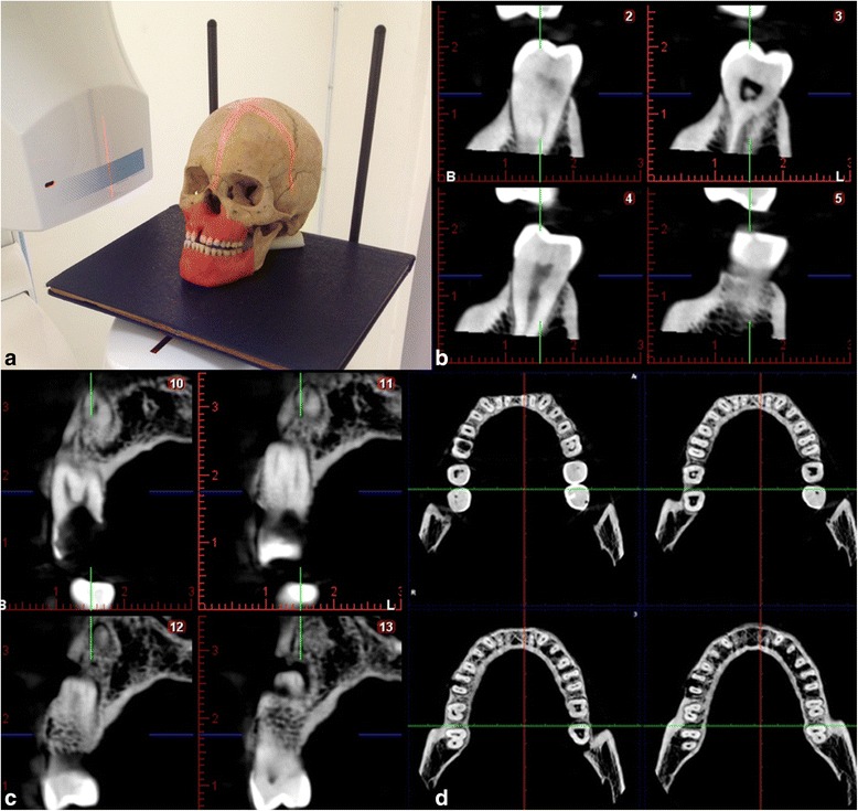
The study material comprised 12 dry skulls with maxilla and mandible. Artificial defects (dehiscence, tunnel, and fenestration) were created on anterior, premolar and molar teeth separately using burs. In total 14 dehiscences, 13 fenestrations, eight tunnel and 16 without periodontal defect were used in the study. These were randomly created on dry skulls. Each teeth with and without defects were images at various vertical angles using each of the following modalities: a Planmeca Promax Cone Beam CT and a Digora photostimulable phosphor plates. Specificity and sensitivity for assessing periodontal defects by each radiographic technique were calculated. Chi-square statistics were used to evaluate differences between modalities. Kappa statistics assessed the agreement between observers. Results were considered significant at P < 0.05.

RESULTS

The kappa values for inter-observer agreement between observers ranged between 0.78 and 0.96 for the CBCT, and 0.43 and 0.72 of intraoral images. The Kappa values for detecting defects on anterior teeth was the least, following premolar and molar teeth both CBCT and intraoral imaging.

CONCLUSIONS

CBCT has the highest sensitivity and diagnostic accuracy for detecting various periodontal defects among the radiographic modalities examined.

摘要

背景

本研究旨在比较锥形束计算机断层扫描(CBCT)设备与数字化口腔内放射成像技术在检测牙周缺损方面的诊断准确性。

方法

研究材料包括12个带有上颌骨和下颌骨的干燥颅骨。使用牙钻分别在前牙、前磨牙和磨牙上制造人工缺损(骨开裂、骨隧道和骨开窗)。本研究共使用了14处骨开裂、13处骨开窗、8处骨隧道以及16处无牙周缺损的样本。这些样本随机设置在干燥颅骨上。使用以下每种方式在不同垂直角度对有缺损和无缺损的牙齿进行成像:Planmeca Promax锥形束CT和Digora光激励荧光板。计算每种放射成像技术评估牙周缺损的特异性和敏感性。采用卡方统计评估不同方式之间的差异。kappa统计评估观察者之间的一致性。P < 0.05时结果被认为具有显著性。

结果

CBCT观察者间一致性的kappa值在0.78至0.96之间,口腔内图像的kappa值在0.43至0.72之间。CBCT和口腔内成像检测前牙缺损的kappa值最低,其次是前磨牙和磨牙。

结论

在本研究的放射成像方式中,CBCT检测各种牙周缺损的敏感性和诊断准确性最高。

https://cdn.ncbi.nlm.nih.gov/pmc/blobs/6191/4446848/76a106d1a475/12903_2015_46_Fig4_HTML.jpg
https://cdn.ncbi.nlm.nih.gov/pmc/blobs/6191/4446848/db427f50aa95/12903_2015_46_Fig1_HTML.jpg
https://cdn.ncbi.nlm.nih.gov/pmc/blobs/6191/4446848/a1cd47799f21/12903_2015_46_Fig2_HTML.jpg
https://cdn.ncbi.nlm.nih.gov/pmc/blobs/6191/4446848/ab56d43ce463/12903_2015_46_Fig3_HTML.jpg
https://cdn.ncbi.nlm.nih.gov/pmc/blobs/6191/4446848/76a106d1a475/12903_2015_46_Fig4_HTML.jpg

相似文献

1
Comparison of intraoral radiography and cone-beam computed tomography for the detection of periodontal defects: an in vitro study.口腔内放射摄影术与锥形束计算机断层扫描术在检测牙周缺损方面的比较:一项体外研究。
BMC Oral Health. 2015 May 28;15:64. doi: 10.1186/s12903-015-0046-2.
2
Detection of periodontal bone loss using digital intraoral and cone beam computed tomography images: an in vitro assessment of bony and/or infrabony defects.使用数字化口腔内和锥形束计算机断层扫描图像检测牙周骨丧失:对骨和/或骨下缺损的体外评估
Dentomaxillofac Radiol. 2008 Jul;37(5):252-60. doi: 10.1259/dmfr/57711133.
3
Comparison of CBCT with different voxel sizes and intraoral scanner for detection of periodontal defects: an in vitro study.不同体素尺寸锥形束 CT 与口内扫描仪检测牙周缺损的比较:一项体外研究。
Dentomaxillofac Radiol. 2020 Jul;49(5):20190197. doi: 10.1259/dmfr.20190197. Epub 2020 Mar 10.
4
Comparison of the influence of FOV sizes and different voxel resolutions for the assessment of periodontal defects.视野大小和不同体素分辨率对牙周缺损评估的影响比较。
Dentomaxillofac Radiol. 2015;44(7):20150070. doi: 10.1259/dmfr.20150070. Epub 2015 Apr 22.
5
Effectiveness of limited cone-beam computed tomography in the detection of horizontal root fracture.有限锥形束计算机断层扫描在检测水平根折中的有效性
Dent Traumatol. 2009 Jun;25(3):256-61. doi: 10.1111/j.1600-9657.2009.00770.x.
6
Accuracy of cone-beam computed tomography in detecting alveolar bone dehiscences and fenestrations.锥形束计算机断层扫描在检测牙槽骨骨缺损和骨开窗方面的准确性。
Am J Orthod Dentofacial Orthop. 2015 Mar;147(3):313-23. doi: 10.1016/j.ajodo.2014.10.032.
7
Assessment of buccal marginal alveolar peri-implant and periodontal defects using a cone beam CT system with and without the application of metal artefact reduction mode.使用配备和不配备金属伪影降低模式的锥形束 CT 系统评估颊侧边缘牙槽嵴种植体和牙周缺损。
Dentomaxillofac Radiol. 2013;42(8):20130176. doi: 10.1259/dmfr.20130176.
8
Observer performance based on marginal bone tissue visibility in Scanora panoramic radiography and posterior bitewing radiography.基于Scanora全景X线摄影和后牙咬合翼片X线摄影中边缘骨组织可见性的观察者表现。
Stomatologija. 2008;10(1):36-43.
9
Digital method for quantification of circumferential periodontal bone level using cone beam CT.利用锥形束 CT 进行牙周骨水平周径定量的数字化方法。
Clin Oral Investig. 2013 Mar;17(2):389-96. doi: 10.1007/s00784-012-0715-3. Epub 2012 Mar 21.
10
Comparison of intraoral digital radiography and cone-beam computed tomography in the measurement of periodontal bone defects.口腔内数字射线摄影与锥形束 CT 在牙周骨缺损测量中的比较。
Dent Med Probl. 2020 Jul-Sep;57(3):269-273. doi: 10.17219/dmp/118749.

引用本文的文献

1
[Reproducibility and criterion validity of two radiographic techniques for variations in mandibular premolar compared with CBCT techniqueReprodutibilidade e validade do critério de duas técnicas radiográficas para variações dos pré-molares inferiores em relação à TCFC].与锥形束计算机断层扫描(CBCT)技术相比,两种用于下颌前磨牙变异的放射成像技术的可重复性和标准效度[两种放射成像技术用于下颌前磨牙变异相对于CBCT技术的可重复性和标准效度]
Rev Cuid. 2022 Aug 28;13(1):e19. doi: 10.15649/cuidarte.2300. eCollection 2022 Jan-Apr.
2
Evaluation of Osteocalcin Level in Gingival Crevicular Fluid in Periodontal Intrabony Defects Treated with Autologous Platelet Rich Fibrin: Non-Randomized Experimental Study.富血小板纤维蛋白治疗牙周骨内缺损时龈沟液中骨钙素水平的评估:非随机实验研究
J Oral Maxillofac Res. 2024 Dec 31;15(4):e3. doi: 10.5037/jomr.2024.15403. eCollection 2024 Oct-Dec.
3

本文引用的文献

1
CBCT-based volume of simulated root resorption - influence of FOV and voxel size.基于锥形束计算机断层扫描(CBCT)的模拟牙根吸收体积——视野(FOV)和体素大小的影响
Int Endod J. 2015 Oct;48(10):959-65. doi: 10.1111/iej.12390. Epub 2014 Oct 24.
2
Diagnostic accuracy of CBCT for periodontal lesions.锥形束计算机断层扫描(CBCT)对牙周病变的诊断准确性
Clin Oral Investig. 2014 May;18(4):1229-1236. doi: 10.1007/s00784-013-1106-0. Epub 2013 Sep 19.
3
Applications of CBCT in dental practice: a review of the literature.锥形束计算机断层扫描在牙科实践中的应用:文献综述
Challenges in detecting various peri-implant bone defects on modified intraoral oblique radiographic projections: evaluation of an artificial mandibular model.在改良口腔内斜位投照影像上检测各种种植体周围骨缺损的挑战:人工下颌骨模型的评估
Oral Radiol. 2025 Apr;41(2):248-259. doi: 10.1007/s11282-024-00798-8. Epub 2025 Jan 14.
4
Efficacy of two radiographic algorithms for detection of peri-implant bone defects on cone-beam computed tomography scans.两种影像学算法在锥形束计算机断层扫描上检测种植体周围骨缺损的效能
BMC Oral Health. 2025 Jan 7;25(1):39. doi: 10.1186/s12903-024-05397-x.
5
Evaluation of Alveolar Bone Destruction Patterns in the Posterior Region of the Maxilla Through Cone Beam Computer Tomography on 361 Consecutive Patients: Effect of Age and Gender.通过对 361 例连续患者的锥形束计算机断层扫描评估上颌后区牙槽骨破坏模式:年龄和性别影响。
Clin Exp Dent Res. 2024 Dec;10(6):e70000. doi: 10.1002/cre2.70000.
6
Skeletal effects of posterior crossbite treatment with either quad helix or rapid maxillary expansion: a randomized controlled trial with 1-year follow-up.Quad helix 或快速扩弓治疗后牙反(牙合)的骨骼效应:一项为期 1 年随访的随机对照试验。
Angle Orthod. 2024 Sep 1;94(5):512-521. doi: 10.2319/010424-9.1.
7
Evaluation of the accuracy of cone beam computed tomography (CBCT) in the detection of peri-implant fenestration.评估锥形束计算机断层扫描(CBCT)在检测种植体周围开窗中的准确性。
BMC Oral Health. 2024 Aug 9;24(1):922. doi: 10.1186/s12903-024-04674-z.
8
Cone-Beam Computed Tomography (CBCT)-Based Diagnosis of Dental Bone Defects.基于锥形束计算机断层扫描(CBCT)的牙骨缺损诊断
Diagnostics (Basel). 2024 Jul 1;14(13):1404. doi: 10.3390/diagnostics14131404.
9
Development and validation of a clinical phantom reproducing various lesions for oral and maxillofacial radiology research.用于口腔颌面放射学研究的、可再现各种病变的临床体模的开发与验证。
Imaging Sci Dent. 2023 Dec;53(4):345-353. doi: 10.5624/isd.20230129. Epub 2023 Sep 25.
10
Comparative Assessment of Reliability and Accuracy of Cone-Beam Computed Tomography (CBCT) Over Direct Surgical Measurement for Periodontal Bone Loss: A Prospective, Cross-Sectional Study.锥形束计算机断层扫描(CBCT)与牙周骨丧失直接手术测量的可靠性和准确性比较评估:一项前瞻性横断面研究。
Cureus. 2023 Sep 3;15(9):e44608. doi: 10.7759/cureus.44608. eCollection 2023 Sep.
Gen Dent. 2012 Sep-Oct;60(5):390-400; quiz 401-2.
4
Validity of wax and acrylic as soft-tissue simulation materials used in in vitro radiographic studies.蜡和丙烯酸酯作为软组织模拟材料在体外放射学研究中的有效性。
Dentomaxillofac Radiol. 2012 Dec;41(8):686-90. doi: 10.1259/dmfr/33467269. Epub 2012 Aug 29.
5
Detection of simulated incipient furcation involvement by CBCT: an in vitro study using pig mandibles.应用 CBCT 检测下颌骨标本中模拟的牙周联合病变:一项体外研究。
Braz Oral Res. 2012 Jul-Aug;26(4):341-7. doi: 10.1590/s1806-83242012000400010.
6
Digital method for quantification of circumferential periodontal bone level using cone beam CT.利用锥形束 CT 进行牙周骨水平周径定量的数字化方法。
Clin Oral Investig. 2013 Mar;17(2):389-96. doi: 10.1007/s00784-012-0715-3. Epub 2012 Mar 21.
7
Detection of periodontal bone loss using cone beam CT and intraoral radiography.使用锥形束 CT 和口腔内射线照相术检测牙周骨损失。
Dentomaxillofac Radiol. 2012 Jan;41(1):64-9. doi: 10.1259/dmfr/13676777.
8
Artefacts in CBCT: a review.CBCT 中的伪影:综述。
Dentomaxillofac Radiol. 2011 Jul;40(5):265-73. doi: 10.1259/dmfr/30642039.
9
Comparative evaluation among different materials to replace soft tissue in oral radiology studies.不同材料在口腔放射学研究中替代软组织的比较评估。
J Appl Oral Sci. 2010 May-Jun;18(3):264-7. doi: 10.1590/s1678-77572010000300012.
10
Accuracy of three-dimensional imaging in assessing maxillary molar furcation involvement.三维成像评估上颌磨牙分叉受累的准确性。
J Clin Periodontol. 2010 May;37(5):436-41. doi: 10.1111/j.1600-051X.2010.01556.x. Epub 2010 Mar 23.