Hueper Katja, Gutberlet Marcel, Bräsen Jan Hinrich, Jang Mi-Sun, Thorenz Anja, Chen Rongjun, Hertel Barbara, Barrmeyer Amelie, Schmidbauer Martina, Meier Martin, von Vietinghoff Sibylle, Khalifa Abedalrazag, Hartung Dagmar, Haller Hermann, Wacker Frank, Rong Song, Gueler Faikah
Institute for Diagnostic and Interventional Radiology, Hannover Medical School, Hannover, Germany.
Institute for Pathology, Hannover Medical School, Hannover, Germany.
PLoS One. 2016 Sep 15;11(9):e0162705. doi: 10.1371/journal.pone.0162705. eCollection 2016.
Kidney transplantation (ktx) in mice is used to learn about rejection and to develop new treatment strategies. Past studies have mainly been based on histological or molecular biological methods. Imaging techniques to monitor allograft pathology have rarely been used.
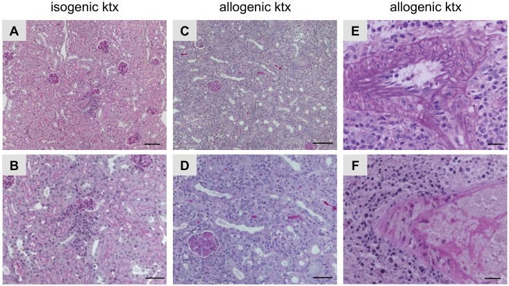
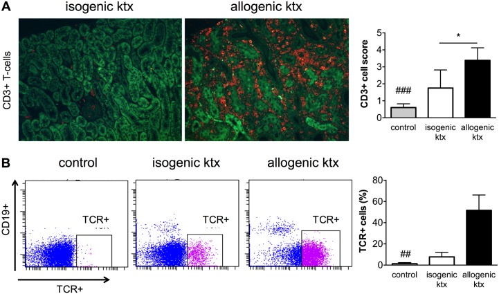
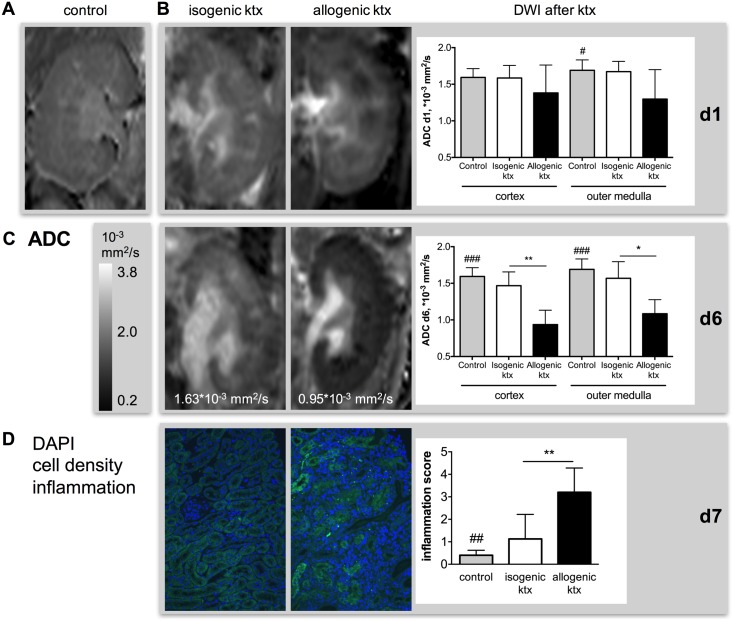
Here we investigated mice after isogenic and allogenic ktx over time with functional MRI with diffusion-weighted imaging (DWI) and mapping of T2-relaxation time (T2-mapping) to assess graft inflammation and edema formation. To characterize graft pathology, we used PAS-staining, counted CD3-positive T-lymphocytes, analyzed leukocytes by means flow cytometry.
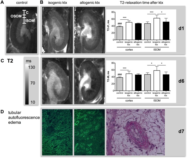
DWI revealed progressive restriction of diffusion of water molecules in allogenic kidney grafts. This was paralleled by enhanced infiltration of the kidney by inflammatory cells. Changes in tissue diffusion were not seen following isogenic ktx. T2-times in renal cortex were increased after both isogenic and allogenic transplantation, consistent with tissue edema due to ischemic injury following prolonged cold ischemia time of 60 minutes. Lack of T2 increase in the inner stripe of the inner medulla in allogenic kidney grafts matched loss of tubular autofluorescence and may result from rejection-driven reductions in tubular water content due to tubular dysfunction and renal functional impairment.
Functional MRI is a valuable non-invasive technique for monitoring inflammation, tissue edema and tubular function. It permits on to differentiate between acute rejection and ischemic renal injury in a mouse model of ktx.
小鼠肾移植用于了解排斥反应并开发新的治疗策略。过去的研究主要基于组织学或分子生物学方法。很少使用成像技术来监测同种异体移植的病理情况。
在此,我们通过功能磁共振成像(functional MRI)结合扩散加权成像(DWI)和T2弛豫时间映射(T2-mapping),对同基因和异基因肾移植后的小鼠进行长期研究,以评估移植物炎症和水肿形成。为了表征移植物病理,我们使用过碘酸雪夫染色(PAS-staining)、计数CD3阳性T淋巴细胞、通过流式细胞术分析白细胞。
DWI显示异基因肾移植中水分子扩散逐渐受限。这与炎性细胞对肾脏的浸润增强相平行。同基因肾移植后未观察到组织扩散的变化。同基因和异基因移植后肾皮质的T2时间均增加,这与60分钟的长时间冷缺血后因缺血性损伤导致的组织水肿一致。异基因肾移植中肾髓质内带T2未增加,与肾小管自发荧光丧失相符,可能是由于肾小管功能障碍和肾功能损害导致排斥反应引起的肾小管含水量降低所致。
功能磁共振成像是监测炎症、组织水肿和肾小管功能的一种有价值的非侵入性技术。它能够在小鼠肾移植模型中区分急性排斥反应和缺血性肾损伤。