Dong Wu, Liu Jie, Wei Lixin, Jingfeng Yang, Chernick Melissa, Hinton David E
Inner Mongolia Provincial Key Laboratory for Toxicants and Animal Disease, College of Animal Science and Technology, Inner Mongolia University for the Nationalities, Tongliao, China; Nicholas School of the Environment, Duke University, Durham, NC, United States.
Zunyi Medical College, Department of Pharmacology , Zunyi , China.
PeerJ. 2016 Aug 23;4:e2282. doi: 10.7717/peerj.2282. eCollection 2016.
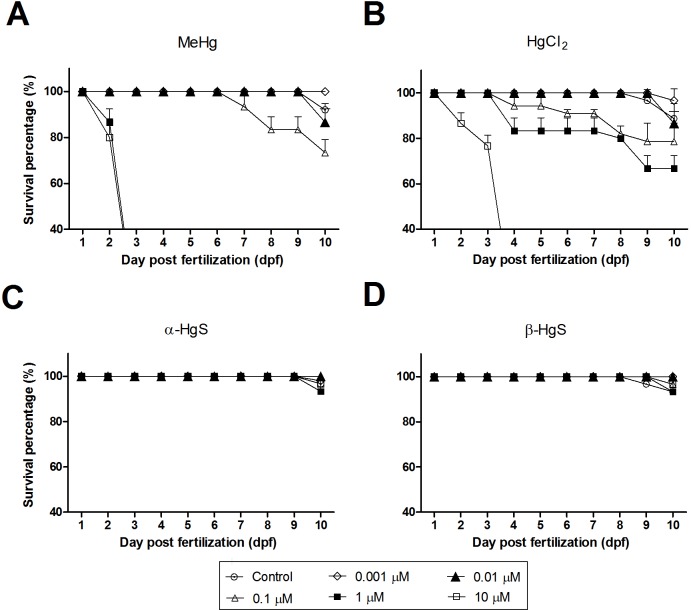
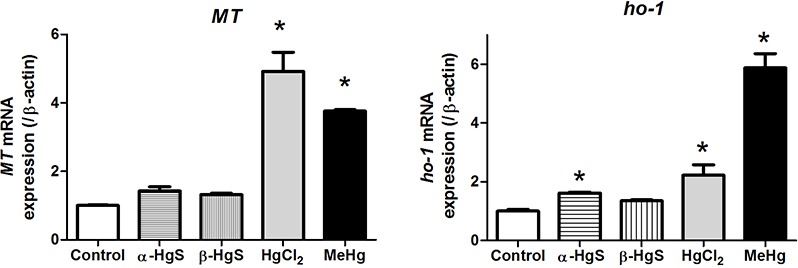
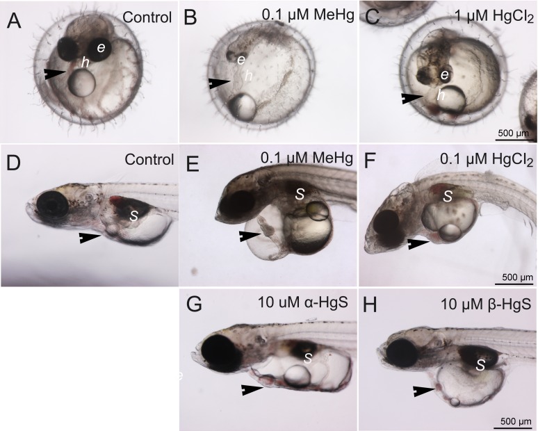
This study examined developmental toxicity of different mercury compounds, including some used in traditional medicines. Medaka (Oryzias latipes) embryos were exposed to 0.001-10 µM concentrations of MeHg, HgCl2, α-HgS (Zhu Sha), and β-HgS (Zuotai) from stage 10 (6-7 hpf) to 10 days post fertilization (dpf). Of the forms of mercury in this study, the organic form (MeHg) proved the most toxic followed by inorganic mercury (HgCl2), both producing embryo developmental toxicity. Altered phenotypes included pericardial edema with elongated or tube heart, reduction of eye pigmentation, and failure of swim bladder inflation. Both α-HgS and β-HgS were less toxic than MeHg and HgCl2. Total RNA was extracted from survivors three days after exposure to MeHg (0.1 µM), HgCl2 (1 µM), α-HgS (10 µM), or β-HgS (10 µM) to examine toxicity-related gene expression. MeHg and HgCl2 markedly induced metallothionein (MT) and heme oxygenase-1 (Ho-1), while α-HgS and β-HgS failed to induce either gene. Chemical forms of mercury compounds proved to be a major determinant in their developmental toxicity.
本研究检测了不同汞化合物的发育毒性,其中包括一些传统药物中使用的汞化合物。将青鳉(Oryzias latipes)胚胎从受精后第10阶段(6 - 7小时胚胎期)至受精后10天暴露于浓度为0.001 - 10 μM的甲基汞、氯化汞、α-硫化汞(朱砂)和β-硫化汞(坐胎)中。在本研究的汞形态中,有机形态(甲基汞)毒性最强,其次是无机汞(氯化汞),二者均产生胚胎发育毒性。改变的表型包括心包水肿伴心脏拉长或呈管状、眼色素沉着减少以及鳔充气失败。α-硫化汞和β-硫化汞的毒性均低于甲基汞和氯化汞。在暴露于甲基汞(0.1 μM)、氯化汞(1 μM)、α-硫化汞(10 μM)或β-硫化汞(10 μM)三天后,从存活胚胎中提取总RNA,以检测与毒性相关的基因表达。甲基汞和氯化汞显著诱导金属硫蛋白(MT)和血红素加氧酶-1(Ho-1),而α-硫化汞和β-硫化汞未能诱导这两种基因。汞化合物的化学形态被证明是其发育毒性的主要决定因素。