Petit C, Gouel F, Dubus I, Heuclin C, Roget K, Vannier J P
MERCI EA3829, Université de Rouen, Faculté de Médecine-Pharmacie, 22 Boulevard Gambetta, 76183, Rouen, France.
BioSIMS Technologies, 75 Route de Lyons la forêt, Seine BioPolis, 76000, Rouen, France.
BMC Cancer. 2016 Sep 22;16(1):746. doi: 10.1186/s12885-016-2776-1.
Several studies show that bone marrow (BM) microenvironment and hypoxia condition can promote the survival of leukemic cells and induce resistance to anti-leukemic drugs. However, the molecular mechanism for chemoresistance by hypoxia is not fully understood.
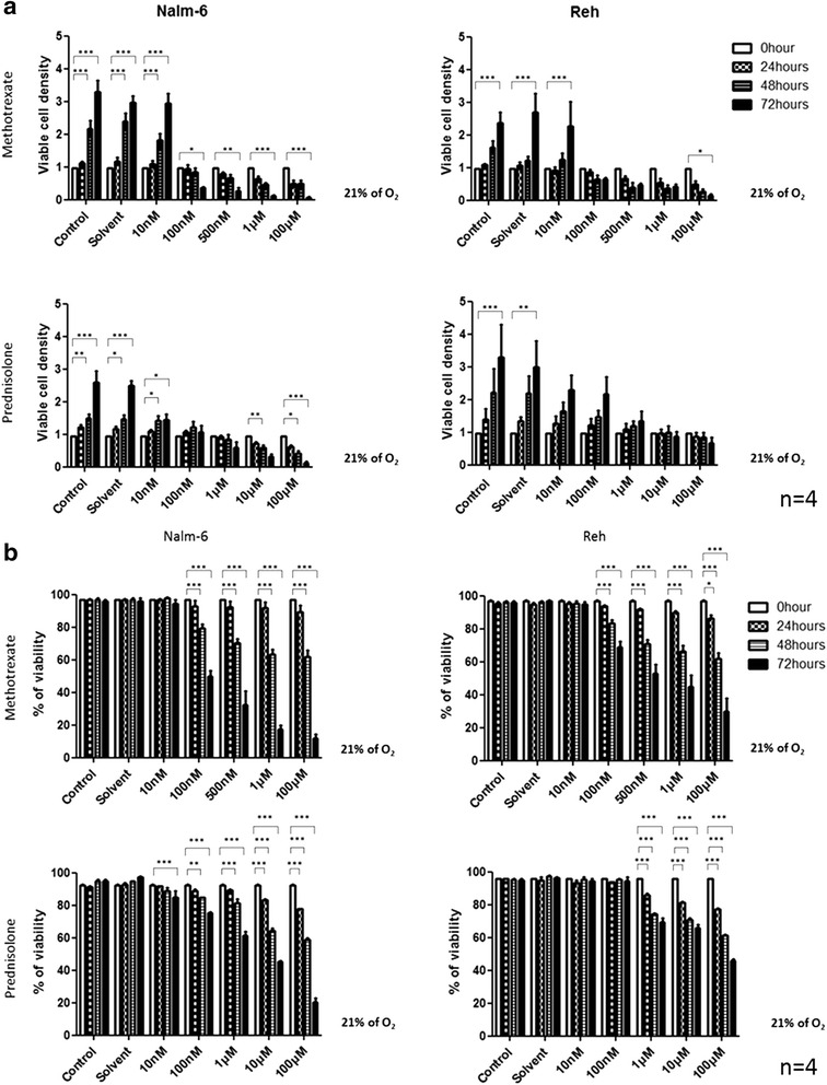
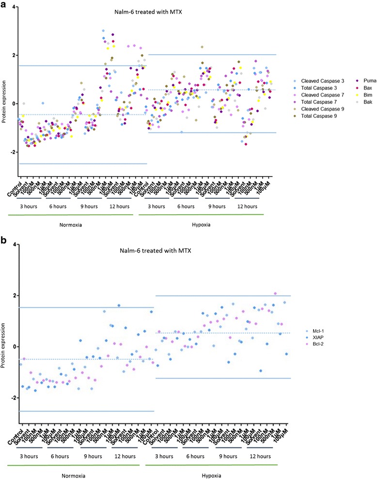
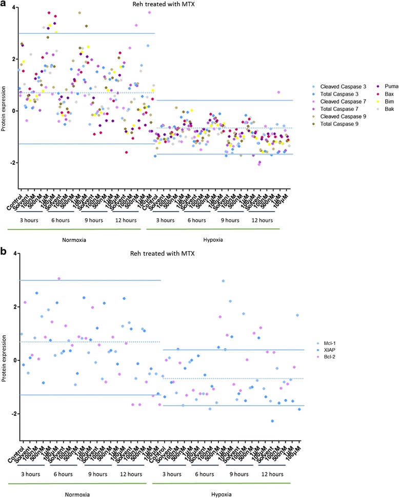
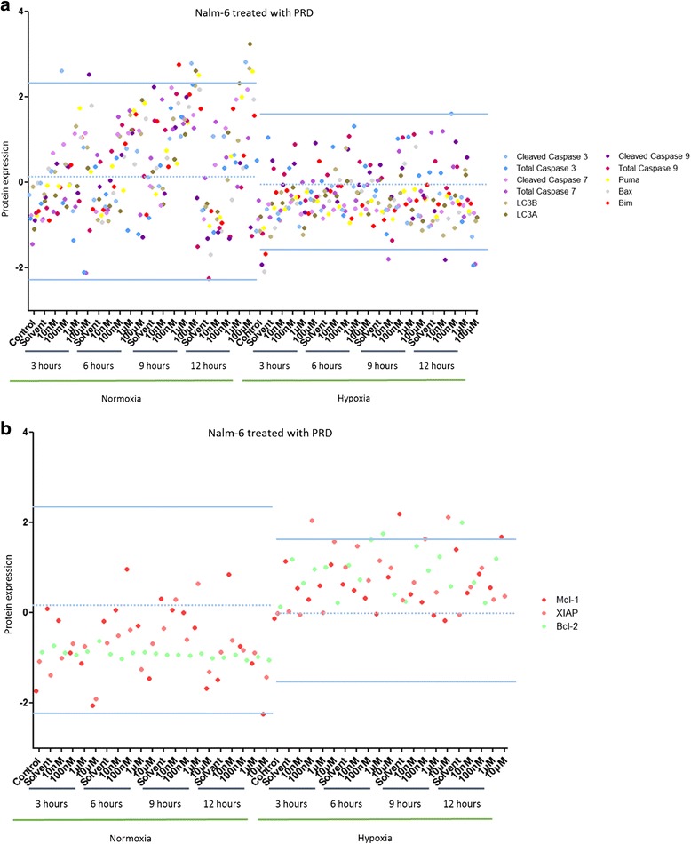
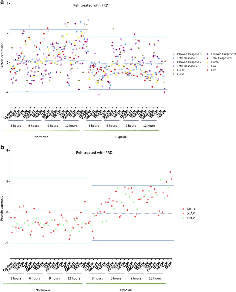
In the present study, we investigated the effect of hypoxia on resistance to two therapies, methotrexate (MTX) and prednisolone (PRD), in two cell models for acute lymphoblastic leukemia (ALL). To look for an implication of hypoxia in chemoresistance, cell viability, total cell density and cell proliferation were analyzed. Survival and death signaling pathways were also screened by "reverse phase protein array" (RPPA) and western blotting experiments conducted on selected proteins to confirm the results.
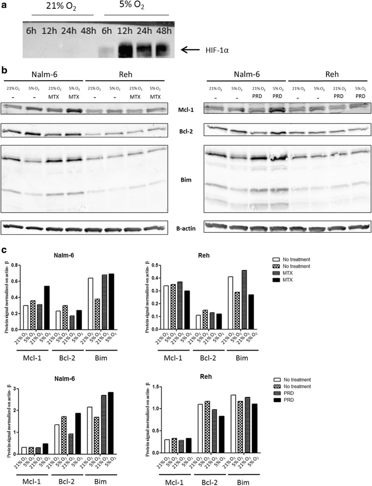
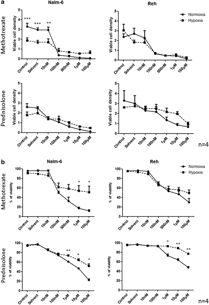
We found that hypoxia promotes chemoresistance in both ALL cell lines. The induction of drug-resistance by hypoxia was not associated with an increase in total cell density nor an increase in cell proliferation. Using RPPA, we show that chemoresistance induced by hypoxia was mediated through an alteration of cell death signaling pathways. This protective effect of hypoxia seems to occur via a decrease in pro-apoptotic proteins and an increase in anti-apoptotic proteins. The results were confirmed by immunoblotting. Indeed, hypoxia is able to modulate the expression of anti-apoptotic proteins independently of chemotherapy while a pro-apoptotic signal induced by a chemotherapy is not modulated by hypoxia.
Hypoxia is a factor in leukemia cell resistance and for two conventional chemotherapies modulates cell death signaling pathways without affecting total cell density or cell proliferation.
多项研究表明,骨髓(BM)微环境和缺氧条件可促进白血病细胞存活并诱导其对抗白血病药物产生耐药性。然而,缺氧导致化疗耐药的分子机制尚未完全明确。
在本研究中,我们在两种急性淋巴细胞白血病(ALL)细胞模型中研究了缺氧对甲氨蝶呤(MTX)和泼尼松龙(PRD)两种治疗方法耐药性的影响。为探究缺氧在化疗耐药中的作用,我们分析了细胞活力、总细胞密度和细胞增殖情况。还通过“反向蛋白质阵列”(RPPA)以及对选定蛋白质进行的蛋白质印迹实验筛选了生存和死亡信号通路,以证实结果。
我们发现缺氧可促进两种ALL细胞系的化疗耐药性。缺氧诱导的耐药性与总细胞密度增加或细胞增殖增加无关。使用RPPA,我们表明缺氧诱导的化疗耐药性是通过细胞死亡信号通路的改变介导的。缺氧的这种保护作用似乎是通过促凋亡蛋白减少和抗凋亡蛋白增加而发生的。蛋白质印迹证实了结果。事实上,缺氧能够独立于化疗调节抗凋亡蛋白的表达,而化疗诱导的促凋亡信号不受缺氧调节。
缺氧是白血病细胞耐药的一个因素,对于两种传统化疗方法,它可调节细胞死亡信号通路,而不影响总细胞密度或细胞增殖。