Hortemo Kristin Halvorsen, Lunde Per Kristian, Anonsen Jan Haug, Kvaløy Heidi, Munkvik Morten, Rehn Tommy Aune, Sjaastad Ivar, Lunde Ida Gjervold, Aronsen Jan Magnus, Sejersted Ole M
Institute for Experimental Medical Research, Oslo University Hospital and University of Oslo, Oslo, Norway Center for Heart Failure Research, University of Oslo, Oslo, Norway
Institute for Experimental Medical Research, Oslo University Hospital and University of Oslo, Oslo, Norway Center for Heart Failure Research, University of Oslo, Oslo, Norway.
Physiol Rep. 2016 Sep;4(18). doi: 10.14814/phy2.12896.
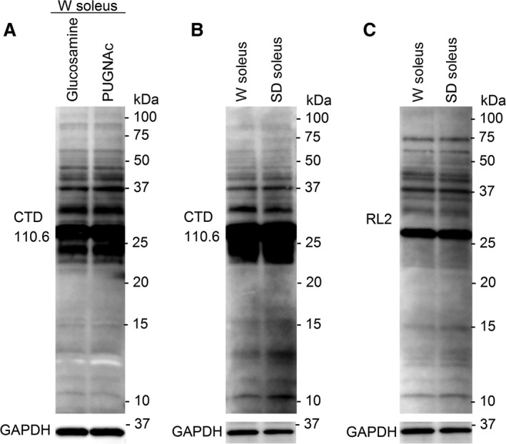
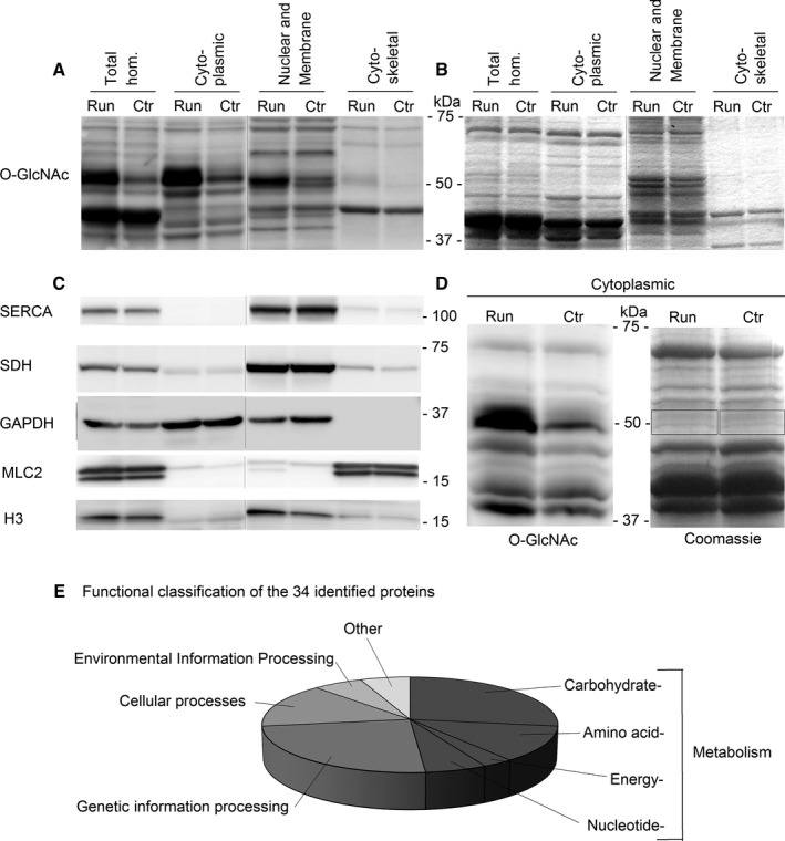
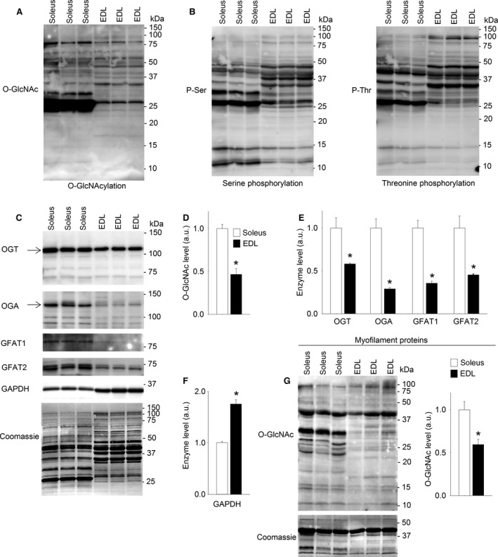
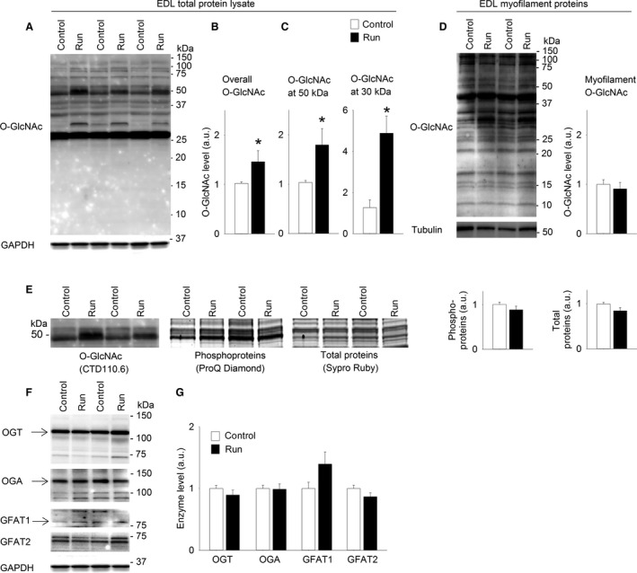
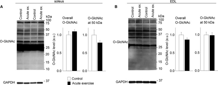
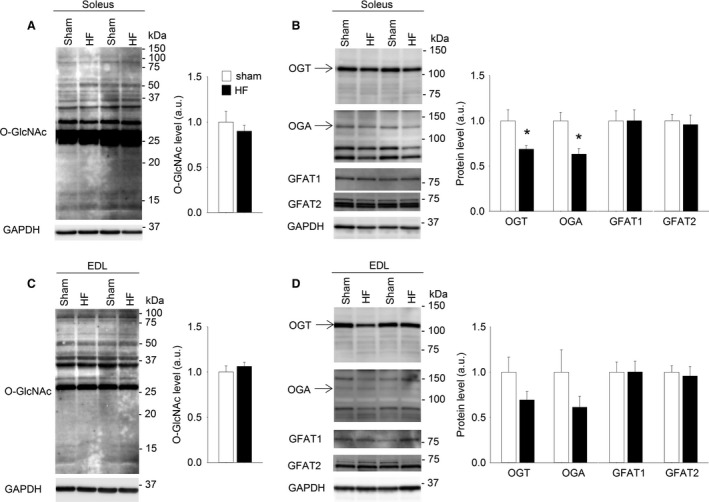
Protein O-GlcNAcylation has emerged as an important intracellular signaling system with both physiological and pathophysiological functions, but the role of protein O-GlcNAcylation in skeletal muscle remains elusive. In this study, we tested the hypothesis that protein O-GlcNAcylation is a dynamic signaling system in skeletal muscle in exercise and disease. Immunoblotting showed different protein O-GlcNAcylation pattern in the prototypical slow twitch soleus muscle compared to fast twitch EDL from rats, with greater O-GlcNAcylation level in soleus associated with higher expression of the modulating enzymes O-GlcNAc transferase (OGT), O-GlcNAcase (OGA), and glutamine fructose-6-phosphate amidotransferase isoforms 1 and 2 (GFAT1, GFAT2). Six weeks of exercise training by treadmill running, but not an acute exercise bout, increased protein O-GlcNAcylation in rat soleus and EDL There was a striking increase in O-GlcNAcylation of cytoplasmic proteins ~50 kDa in size that judged from mass spectrometry analysis could represent O-GlcNAcylation of one or more key metabolic enzymes. This suggests that cytoplasmic O-GlcNAc signaling is part of the training response. In contrast to exercise training, postinfarction heart failure (HF) in rats and humans did not affect skeletal muscle O-GlcNAcylation level, indicating that aberrant O-GlcNAcylation cannot explain the skeletal muscle dysfunction in HF Human skeletal muscle displayed extensive protein O-GlcNAcylation that by large mirrored the fiber-type-related O-GlcNAcylation pattern in rats, suggesting O-GlcNAcylation as an important signaling system also in human skeletal muscle.
蛋白质O-连接的N-乙酰葡糖胺化已成为一种具有生理和病理生理功能的重要细胞内信号系统,但蛋白质O-连接的N-乙酰葡糖胺化在骨骼肌中的作用仍不清楚。在本研究中,我们验证了蛋白质O-连接的N-乙酰葡糖胺化是运动和疾病状态下骨骼肌中一种动态信号系统的假说。免疫印迹显示,与大鼠快肌趾长伸肌(EDL)相比,典型的慢肌比目鱼肌中蛋白质O-连接的N-乙酰葡糖胺化模式不同,比目鱼肌中更高的O-连接的N-乙酰葡糖胺化水平与调节酶O-连接的N-乙酰葡糖胺转移酶(OGT)、O-连接的N-乙酰葡糖胺酶(OGA)以及谷氨酰胺果糖-6-磷酸酰胺转移酶同工型1和2(GFAT1、GFAT2)的更高表达相关。通过跑步机跑步进行六周的运动训练,而非一次急性运动 bout,可增加大鼠比目鱼肌和EDL中的蛋白质O-连接的N-乙酰葡糖胺化。质谱分析判断,大小约为50 kDa的细胞质蛋白的O-连接的N-乙酰葡糖胺化显著增加,可能代表一种或多种关键代谢酶的O-连接的N-乙酰葡糖胺化。这表明细胞质O-连接的N-乙酰葡糖胺信号是训练反应的一部分。与运动训练相反,大鼠和人类的心肌梗死后心力衰竭(HF)并未影响骨骼肌的O-连接的N-乙酰葡糖胺化水平,表明异常的O-连接的N-乙酰葡糖胺化无法解释HF中的骨骼肌功能障碍。人类骨骼肌显示出广泛的蛋白质O-连接的N-乙酰葡糖胺化,这在很大程度上反映了大鼠中与纤维类型相关的O-连接的N-乙酰葡糖胺化模式,表明O-连接的N-乙酰葡糖胺化也是人类骨骼肌中的一种重要信号系统。