Kamran Hassan, Nambi Vijay, Negi Smita, Yang Eric Y, Chen Changyi, Virani Salim S, Kougias Panos, Lumsden Alan B, Morrisett Joel D, Ballantyne Christie M, Brunner Gerd
Division of Atherosclerosis and Vascular Medicine, Department of Medicine, Baylor College of Medicine, Houston, Texas.
Michael E DeBakey VA Medical Center, Houston, Texas; Division of Atherosclerosis and Vascular Medicine, Department of Medicine, Baylor College of Medicine, Houston, Texas; Methodist DeBakey Heart and Vascular Center, Houston Methodist Hospital, Houston, Texas.
Am J Cardiol. 2016 Nov 1;118(9):1399-1404. doi: 10.1016/j.amjcard.2016.07.051. Epub 2016 Aug 13.
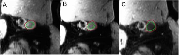
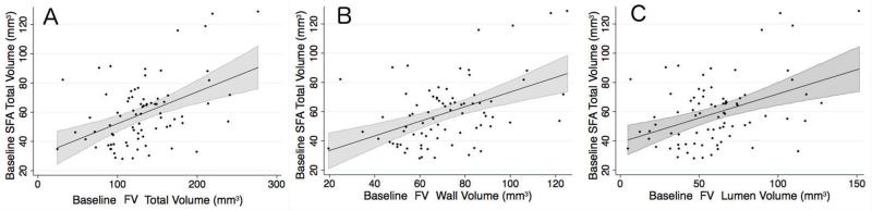
The relation between the arterial and venous systems in patients with impaired lower extremity blood flow remains poorly described. The objective of this secondary analysis of the Effectiveness of Intensive Lipid Modification Medication in Preventing the Progression on Peripheral Artery Disease Trial was to determine the association between femoral vein (FV) volumes and measurements of peripheral artery disease. FV wall, lumen, and total volumes were quantified with fast spin-echo proton density-weighted magnetic resonance imaging scans in 79 patients with peripheral artery disease over 2 years. Reproducibility was excellent for FV total vessel (intraclass correlation coefficient 0.924, confidence interval 0.910 to 0.935) and lumen volumes (intraclass correlation coefficient 0.893, confidence interval 0.873 to 0.910). Baseline superficial femoral artery volumes were directly associated with FV wall (r = 0.46, p <0.0001), lumen (r = 0.42, p = 0.0001), and total volumes (r = 0.46, p <0.0001). The 2-year change in maximum walking time was inversely associated with the 24-month change in FV total volume (r = -0.45, p = 0.03). In conclusion, FV volumes can be measured reliably with fast spin-echo proton density-weighted magnetic resonance imaging, and baseline superficial femoral artery plaque burden is positively associated with FV volumes, whereas the 2-year change in FV volumes and leg function show an inverse relation.
下肢血流受损患者的动静脉系统之间的关系仍未得到充分描述。这项对强化脂质修饰药物预防外周动脉疾病进展试验的二次分析的目的是确定股静脉(FV)容积与外周动脉疾病测量值之间的关联。在2年时间里,对79例外周动脉疾病患者进行快速自旋回波质子密度加权磁共振成像扫描,以量化FV壁、管腔和总体积。FV总血管(组内相关系数0.924,置信区间0.910至0.935)和管腔容积(组内相关系数0.893,置信区间0.873至0.910)的可重复性极佳。基线股浅动脉容积与FV壁(r = 0.46,p <0.0001)、管腔(r = 0.42,p = 0.0001)和总体积(r = 0.46,p <0.0001)直接相关。最大步行时间的2年变化与FV总体积的24个月变化呈负相关(r = -0.45,p = 0.03)。总之,使用快速自旋回波质子密度加权磁共振成像能够可靠地测量FV容积,基线股浅动脉斑块负荷与FV容积呈正相关,而FV容积的2年变化与腿部功能呈负相关。