• 文献检索
  • 文档翻译
  • 深度研究
  • 学术资讯
  • Suppr Zotero 插件Zotero 插件
  • 邀请有礼
  • 套餐&价格
  • 历史记录
应用&插件
Suppr Zotero 插件Zotero 插件浏览器插件Mac 客户端Windows 客户端微信小程序
定价
高级版会员购买积分包购买API积分包
服务
文献检索文档翻译深度研究API 文档MCP 服务
关于我们
关于 Suppr公司介绍联系我们用户协议隐私条款
关注我们

Suppr 超能文献

核心技术专利:CN118964589B侵权必究
粤ICP备2023148730 号-1Suppr @ 2026

文献检索

告别复杂PubMed语法,用中文像聊天一样搜索,搜遍4000万医学文献。AI智能推荐,让科研检索更轻松。

立即免费搜索

文件翻译

保留排版,准确专业,支持PDF/Word/PPT等文件格式,支持 12+语言互译。

免费翻译文档

深度研究

AI帮你快速写综述,25分钟生成高质量综述,智能提取关键信息,辅助科研写作。

立即免费体验

间充质基质细胞片对屈肌腱愈合炎症阶段的影响。

The effect of mesenchymal stromal cell sheets on the inflammatory stage of flexor tendon healing.

作者信息

Shen Hua, Kormpakis Ioannis, Havlioglu Necat, Linderman Stephen W, Sakiyama-Elbert Shelly E, Erickson Isaac E, Zarembinski Thomas, Silva Matthew J, Gelberman Richard H, Thomopoulos Stavros

机构信息

Department of Orthopaedic Surgery, Washington University, 660 South Euclid, Campus, Box 8233, St. Louis, MO, 63110, USA.

Department of Pathology, John Cochran VA Medical Center, St. Louis, MO, USA.

出版信息

Stem Cell Res Ther. 2016 Sep 27;7(1):144. doi: 10.1186/s13287-016-0406-0.

DOI:10.1186/s13287-016-0406-0
PMID:27677963
原文链接:https://pmc.ncbi.nlm.nih.gov/articles/PMC5039894/
Abstract

BACKGROUND

The clinical outcomes following intrasynovial flexor tendon repair are highly variable. Excessive inflammation is a principal factor underlying the formation of adhesions at the repair surface and affecting matrix regeneration at the repair center that limit tendon excursion and impair tendon healing. A previous in-vitro study revealed that adipose-derived mesenchymal stromal cells (ASCs) modulate tendon fibroblast response to macrophage-induced inflammation. The goal of the current study was therefore to explore the effectiveness of autologous ASCs on the inflammatory stage of intrasynovial tendon healing in vivo using a clinically relevant animal model.

METHODS

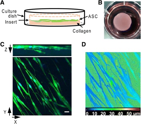
Zone II flexor tendon transections and suture repairs were performed in a canine model. Autologous ASC sheets were delivered to the surface of repaired tendons. Seven days after repair, the effects of ASCs on tendon healing, with a focus on the inflammatory response, were evaluated using gene expression assays, immunostaining, and histological assessments.

RESULTS

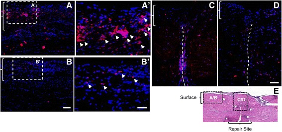
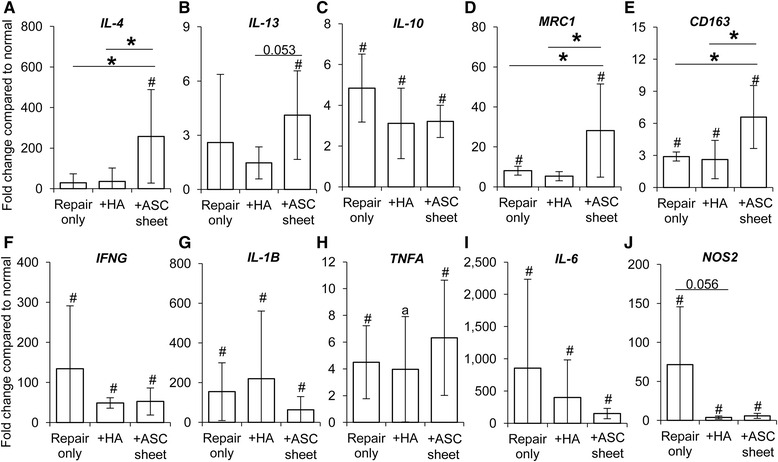
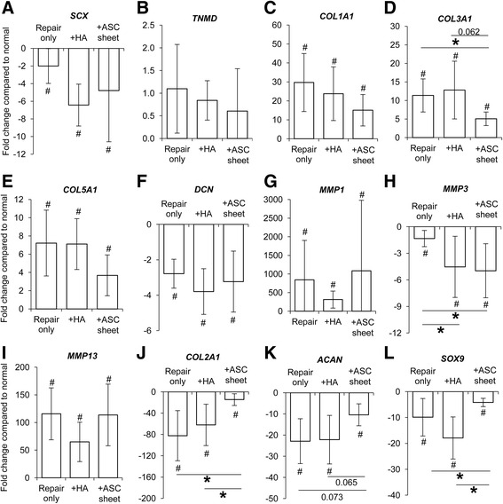
ASCs delivered via the cell sheet infiltrated the host tendon, including the repair surface and the space between the tendon ends, as viewed histologically by tracking GFP-expressing ASCs. Gene expression results demonstrated that ASCs promoted a regenerative/anti-inflammatory M2 macrophage phenotype and regulated tendon matrix remodeling. Specifically, there were significant increases in M2-stimulator (IL-4), marker (CD163 and MRC1), and effector (VEGF) gene expression in ASC-sheet treated tendons compared with nontreated tendons. When examining changes in extracellular matrix expression, tendon injury caused a significant increase in scar-associated COL3A1 expression and reductions in COL2A1 and ACAN expression. The ASC treatment effectively counteracted these changes, returning the expression levels of these genes closer to normal. Immunostaining further confirmed that ASC treatment increased CD163 M2 cells in the repaired tendons and suppressed cell apoptosis at the repair site.

CONCLUSIONS

This study provides a novel approach for delivering ASCs with outcomes indicating potential for substantial modulation of the inflammatory environment and enhancement of tendon healing after flexor tendon repair.

摘要

背景

滑膜内屈肌腱修复后的临床结果差异很大。过度炎症是修复表面形成粘连以及影响修复中心基质再生的主要因素,这会限制肌腱的活动并损害肌腱愈合。先前的一项体外研究表明,脂肪来源的间充质基质细胞(ASC)可调节肌腱成纤维细胞对巨噬细胞诱导的炎症的反应。因此,本研究的目的是使用具有临床相关性的动物模型,在体内探索自体ASC对滑膜内肌腱愈合炎症阶段的有效性。

方法

在犬模型中进行II区屈肌腱横断和缝合修复。将自体ASC片放置于修复肌腱的表面。修复7天后,使用基因表达分析、免疫染色和组织学评估来评估ASC对肌腱愈合的影响,重点是炎症反应。

结果

通过追踪表达绿色荧光蛋白的ASC进行组织学观察发现,经细胞片递送的ASC浸润了宿主肌腱,包括修复表面和肌腱断端之间的间隙。基因表达结果表明,ASC促进了再生/抗炎M2巨噬细胞表型并调节肌腱基质重塑。具体而言,与未处理的肌腱相比,ASC片处理的肌腱中M2刺激因子(IL-4)、标志物(CD163和MRC1)和效应因子(VEGF)的基因表达显著增加。在检查细胞外基质表达的变化时,肌腱损伤导致瘢痕相关的COL3A1表达显著增加,而COL2A1和ACAN表达降低。ASC治疗有效地抵消了这些变化,使这些基因的表达水平更接近正常。免疫染色进一步证实,ASC治疗增加了修复肌腱中CD163 M2细胞的数量,并抑制了修复部位的细胞凋亡。

结论

本研究提供了一种递送ASC的新方法,结果表明其有可能显著调节炎症环境并增强屈肌腱修复后的肌腱愈合。

https://cdn.ncbi.nlm.nih.gov/pmc/blobs/f87a/5039894/a83fb8739b90/13287_2016_406_Fig7_HTML.jpg
https://cdn.ncbi.nlm.nih.gov/pmc/blobs/f87a/5039894/3427579d4d9a/13287_2016_406_Fig1_HTML.jpg
https://cdn.ncbi.nlm.nih.gov/pmc/blobs/f87a/5039894/9b86d338b342/13287_2016_406_Fig2_HTML.jpg
https://cdn.ncbi.nlm.nih.gov/pmc/blobs/f87a/5039894/00396069aea0/13287_2016_406_Fig3_HTML.jpg
https://cdn.ncbi.nlm.nih.gov/pmc/blobs/f87a/5039894/3d83ea7e5e6c/13287_2016_406_Fig4_HTML.jpg
https://cdn.ncbi.nlm.nih.gov/pmc/blobs/f87a/5039894/8484d0be4722/13287_2016_406_Fig5_HTML.jpg
https://cdn.ncbi.nlm.nih.gov/pmc/blobs/f87a/5039894/c9428cf5a714/13287_2016_406_Fig6_HTML.jpg
https://cdn.ncbi.nlm.nih.gov/pmc/blobs/f87a/5039894/a83fb8739b90/13287_2016_406_Fig7_HTML.jpg

相似文献

1
The effect of mesenchymal stromal cell sheets on the inflammatory stage of flexor tendon healing.间充质基质细胞片对屈肌腱愈合炎症阶段的影响。
Stem Cell Res Ther. 2016 Sep 27;7(1):144. doi: 10.1186/s13287-016-0406-0.
2
Combined Administration of ASCs and BMP-12 Promotes an M2 Macrophage Phenotype and Enhances Tendon Healing.脂肪干细胞与骨形态发生蛋白-12联合应用可促进M2巨噬细胞表型并增强肌腱愈合。
Clin Orthop Relat Res. 2017 Sep;475(9):2318-2331. doi: 10.1007/s11999-017-5369-7. Epub 2017 May 1.
3
The effect of adipose-derived stem cell sheets and CTGF on early flexor tendon healing in a canine model.脂肪源干细胞片和 CTGF 对犬早期屈肌腱愈合的影响。
Sci Rep. 2018 Jul 23;8(1):11078. doi: 10.1038/s41598-018-29474-8.
4
Effect of adipose-derived stromal cells and BMP12 on intrasynovial tendon repair: A biomechanical, biochemical, and proteomics study.脂肪来源的间充质干细胞和BMP12对滑膜内肌腱修复的影响:一项生物力学、生物化学和蛋白质组学研究。
J Orthop Res. 2016 Apr;34(4):630-40. doi: 10.1002/jor.23064. Epub 2015 Oct 14.
5
Adipose-derived mesenchymal stromal cells modulate tendon fibroblast responses to macrophage-induced inflammation in vitro.脂肪来源的间充质基质细胞在体外调节肌腱成纤维细胞对巨噬细胞诱导的炎症反应。
Stem Cell Res Ther. 2015 Apr 16;6(1):74. doi: 10.1186/s13287-015-0059-4.
6
Stem cell-derived extracellular vesicles attenuate the early inflammatory response after tendon injury and repair.干细胞衍生的细胞外囊泡可减轻肌腱损伤及修复后的早期炎症反应。
J Orthop Res. 2020 Jan;38(1):117-127. doi: 10.1002/jor.24406. Epub 2019 Jul 26.
7
Human Mesenchymal Stromal Cell Sheets Induce Macrophages Predominantly to an Anti-Inflammatory Phenotype.人骨髓间充质基质细胞片主要诱导巨噬细胞向抗炎表型分化。
Stem Cells Dev. 2018 Jul 1;27(13):922-934. doi: 10.1089/scd.2017.0275.
8
The effects of NF-κB suppression on the early healing response following intrasynovial tendon repair in a canine model.NF-κB 抑制对犬滑膜内肌腱修复后早期愈合反应的影响。
J Orthop Res. 2023 Oct;41(10):2295-2304. doi: 10.1002/jor.25576. Epub 2023 May 4.
9
Adeno-associated virus-2-mediated bFGF gene transfer to digital flexor tendons significantly increases healing strength. an in vivo study.腺相关病毒2介导的碱性成纤维细胞生长因子基因转移至指屈肌腱可显著提高愈合强度。一项体内研究。
J Bone Joint Surg Am. 2008 May;90(5):1078-89. doi: 10.2106/JBJS.F.01188.
10
Effect of connective tissue growth factor delivered via porous sutures on the proliferative stage of intrasynovial tendon repair.通过多孔缝线递送的结缔组织生长因子对滑膜内肌腱修复增殖阶段的影响。
J Orthop Res. 2018 Jul;36(7):2052-2063. doi: 10.1002/jor.23842. Epub 2018 Feb 1.

引用本文的文献

1
Regeneration of Biomechanically Functional Tendon Tissue Following Injection of Uncultured, Autologous, Adipose-Derived Regenerative Cells into Partial Achilles Tendon Defects in Rabbits.将未培养的自体脂肪源性再生细胞注射到兔跟腱部分缺损处后生物力学功能腱组织的再生
Int J Mol Sci. 2025 Jul 16;26(14):6800. doi: 10.3390/ijms26146800.
2
Plantar Fasciitis Pathophysiology and the Potential Role of Mesenchymal Stem Cell-Derived Extracellular Vesicles as Therapy.足底筋膜炎的病理生理学以及间充质干细胞衍生的细胞外囊泡作为治疗手段的潜在作用。
Biomedicines. 2025 Jun 23;13(7):1528. doi: 10.3390/biomedicines13071528.
3
Sustainable Immunomodulatory via Macrophage P2Y12 Inhibition Mediated Bioactive Patche for Peritendinous Antiadhesion.

本文引用的文献

1
Primed Mesenchymal Stem Cells Alter and Improve Rat Medial Collateral Ligament Healing.预处理的间充质干细胞改变并改善大鼠内侧副韧带愈合。
Stem Cell Rev Rep. 2016 Feb;12(1):42-53. doi: 10.1007/s12015-015-9633-5.
2
Effect of adipose-derived stromal cells and BMP12 on intrasynovial tendon repair: A biomechanical, biochemical, and proteomics study.脂肪来源的间充质干细胞和BMP12对滑膜内肌腱修复的影响:一项生物力学、生物化学和蛋白质组学研究。
J Orthop Res. 2016 Apr;34(4):630-40. doi: 10.1002/jor.23064. Epub 2015 Oct 14.
3
Adipose-derived mesenchymal stromal cells modulate tendon fibroblast responses to macrophage-induced inflammation in vitro.
通过巨噬细胞P2Y12抑制介导的生物活性贴片实现可持续免疫调节以预防肌腱周围粘连
Adv Sci (Weinh). 2025 Jan;12(4):e2409128. doi: 10.1002/advs.202409128. Epub 2024 Dec 4.
4
Challenges in tendon-bone healing: emphasizing inflammatory modulation mechanisms and treatment.肌腱-骨愈合的挑战:强调炎症调节机制和治疗。
Front Endocrinol (Lausanne). 2024 Nov 6;15:1485876. doi: 10.3389/fendo.2024.1485876. eCollection 2024.
5
GelMA encapsulating BMSCs-exosomes combined with interference screw or suture anchor promotes tendon-bone healing in a rabbit model.GelMA 包被 BMSCs-exosomes 联合干扰螺钉或缝线锚钉促进兔模型腱骨愈合。
Sci Rep. 2024 Nov 15;14(1):28212. doi: 10.1038/s41598-024-79787-0.
6
Evaluation of Patellar Tendon Structural Changes following Biological Treatments: Secondary Analysis of Double-Blinded Clinical Trial of Bone Marrow Mesenchymal Stromal Cells and Leukocyte-Poor Platelet-Rich Plasma.生物治疗后髌腱结构变化的评估:骨髓间充质基质细胞与少白细胞富血小板血浆双盲临床试验的二次分析
Biomedicines. 2024 Jul 18;12(7):1599. doi: 10.3390/biomedicines12071599.
7
Advances in mesenchymal stem cells therapy for tendinopathies.间充质干细胞治疗肌腱病的研究进展。
Chin J Traumatol. 2024 Jan;27(1):11-17. doi: 10.1016/j.cjtee.2023.11.002. Epub 2023 Nov 11.
8
Treatment of Naturally Occurring Tendon Disease with Allogeneic Multipotent Mesenchymal Stromal Cells: A Randomized, Controlled, Triple-Blinded Pilot Study in Horses.同种异体间充质基质细胞治疗自然发生的肌腱疾病:马的随机、对照、三盲先导研究。
Cells. 2023 Oct 24;12(21):2513. doi: 10.3390/cells12212513.
9
Cell Sheet Technology: An Emerging Approach for Tendon and Ligament Tissue Engineering.细胞片层技术:肌腱和韧带组织工程的新兴方法。
Ann Biomed Eng. 2024 Feb;52(2):141-152. doi: 10.1007/s10439-023-03370-3. Epub 2023 Sep 20.
10
Silver nanoparticles-decorated extracellular matrix graft: fabrication and tendon reconstruction performance.银纳米颗粒修饰的细胞外基质移植物:制备及其肌腱重建性能
Biomater Res. 2023 Sep 14;27(1):85. doi: 10.1186/s40824-023-00428-0.
脂肪来源的间充质基质细胞在体外调节肌腱成纤维细胞对巨噬细胞诱导的炎症反应。
Stem Cell Res Ther. 2015 Apr 16;6(1):74. doi: 10.1186/s13287-015-0059-4.
4
Changes in macrophage phenotype and induction of epithelial-to-mesenchymal transition genes following acute Achilles tenotomy and repair.急性跟腱切断和修复后巨噬细胞表型的变化及上皮-间充质转化基因的诱导。
J Orthop Res. 2014 Jul;32(7):944-51. doi: 10.1002/jor.22624. Epub 2014 Apr 4.
5
The M1 and M2 paradigm of macrophage activation: time for reassessment.巨噬细胞活化的M1和M2范式:是时候重新评估了。
F1000Prime Rep. 2014 Mar 3;6:13. doi: 10.12703/P6-13. eCollection 2014.
6
The early inflammatory response after flexor tendon healing: a gene expression and histological analysis.屈肌腱愈合后早期的炎症反应:基因表达和组织学分析。
J Orthop Res. 2014 May;32(5):645-52. doi: 10.1002/jor.22575. Epub 2014 Jan 24.
7
BMP12 induces tenogenic differentiation of adipose-derived stromal cells.BMP12 诱导脂肪来源的基质细胞向肌腱细胞分化。
PLoS One. 2013 Oct 14;8(10):e77613. doi: 10.1371/journal.pone.0077613. eCollection 2013.
8
Mesenchymal stromal cells: sensors and switchers of inflammation.间质基质细胞:炎症的传感器和开关。
Cell Stem Cell. 2013 Oct 3;13(4):392-402. doi: 10.1016/j.stem.2013.09.006.
9
Review of the adipose derived stem cell secretome.脂肪来源干细胞分泌组的综述。
Biochimie. 2013 Dec;95(12):2222-8. doi: 10.1016/j.biochi.2013.06.001. Epub 2013 Jun 14.
10
Topical administration of orbital fat-derived stem cells promotes corneal tissue regeneration.局部应用眼眶脂肪来源的干细胞可促进角膜组织再生。
Stem Cell Res Ther. 2013 Jun 14;4(3):72. doi: 10.1186/scrt223.