Lin Ko-Jo, Loi Mei-Xue, Lien Gi-Shih, Cheng Chieh-Feng, Pao Hsiang-Yin, Chang Yun-Chuang, Ji Andrea Tung-Qian, Ho Jennifer Hui-Chun
Stem Cell Res Ther. 2013 Jun 14;4(3):72. doi: 10.1186/scrt223.
Topical administration of eye drops is the major route for drug delivery to the cornea. Orbital fat-derived stem cells (OFSCs) possess an in vitro corneal epithelial differentiation capacity. Both the safety and immunomodulatory ability of systemic OFSC transplantation were demonstrated in our previous work. In this study, we investigated the safety, therapeutic effect, and mechanism(s) of topical OFSC administration in an extensive alkali-induced corneal wound.
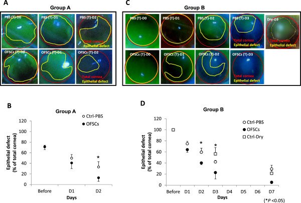
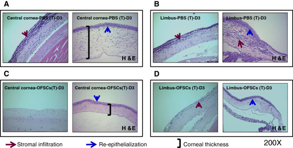
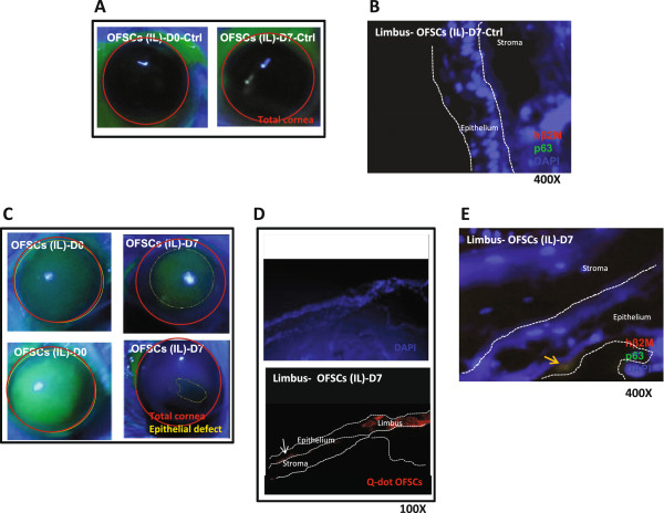
Corneal injury was created by contact of a piece of 0.5 N NaOH-containing filter paper on the corneal surface of a male Balb/c mouse for 30 s. The area of the filter paper covered the central 70% or 100% of the corneal surface. OFSCs (2 × 10(5)) in 5 μl phosphate-buffered saline (PBS) were given by topical administration (T) twice a day or by two intralimbal (IL) injections in the right cornea, while 5 μl of PBS in the left cornea served as the control.
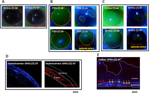
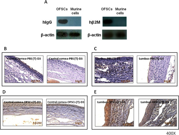
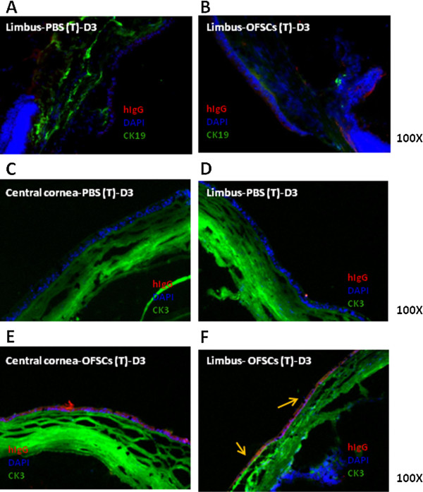
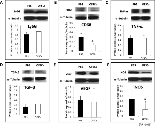
Topical OFSCs promoted corneal re-epithelialization of both the limbal-sparing and limbal-involved corneal wounds. In the first three days, topical OFSCs significantly reduced alkali-induced corneal edema and stromal infiltration according to a histopathological examination. Immunohistochemistry and immunofluorescence staining revealed that transplanted cells were easily detectable in the corneal epithelium, limbal epithelium and stroma, but only some of transplanted cells at the limbal epithelium had differentiated into cytokeratin 3-expressing cells. OFSCs did not alter neutrophil (Ly6G) levels in the cornea, but significantly reduced macrophage (CD68) infiltration and inducible nitrous oxide synthetase (iNOS) production during acute corneal injury as quantified by a Western blot analysis. Continuous topical administration of OFSCs for seven days improved corneal transparency, and this was accompanied by diffuse stromal engraftment of transplanted cells and differentiation into p63-expressing cells at the limbal area. The therapeutic effect of the topical administration of OFSCs was superior to that of the IL injection. OFSCs from the IL injection clustered in the limbal area and central corneal epithelium, which was associated with a persistent corneal haze.
Topical OFSC administration is a simple, non-surgical route for stem cell delivery to promote corneal tissue regeneration through ameliorating acute inflammation and corneal epithelial differentiation. The limbal area serves as a niche for OFSCs differentiating into corneal epithelial cells in the first week, while the stroma is a potential site for anti-inflammation of OFSCs. Inhibition of corneal inflammation is related to corneal transparency.
滴眼剂局部给药是药物递送至角膜的主要途径。眼眶脂肪来源的干细胞(OFSCs)具有体外角膜上皮分化能力。我们之前的研究已证明全身OFSC移植的安全性和免疫调节能力。在本研究中,我们调查了在广泛的碱诱导角膜伤口中局部给予OFSCs的安全性、治疗效果及其机制。
通过将一片含0.5N NaOH的滤纸接触雄性Balb/c小鼠角膜表面30秒造成角膜损伤。滤纸覆盖角膜表面中央70%或100%的区域。将5μl磷酸盐缓冲盐水(PBS)中含有的2×10(5)个OFSCs通过局部给药(T)每天两次或通过在右眼角膜进行两次角膜缘内(IL)注射给药,而左眼角膜给予5μl PBS作为对照。
局部给予OFSCs促进了角膜缘保留和角膜缘受累角膜伤口的角膜再上皮化。在头三天,根据组织病理学检查,局部给予OFSCs显著减轻了碱诱导的角膜水肿和基质浸润。免疫组织化学和免疫荧光染色显示,移植细胞在角膜上皮、角膜缘上皮和基质中易于检测到,但仅角膜缘上皮处的一些移植细胞分化为表达细胞角蛋白3的细胞。OFSCs未改变角膜中的中性粒细胞(Ly6G)水平,但通过蛋白质免疫印迹分析定量显示,在急性角膜损伤期间显著减少了巨噬细胞(CD68)浸润和诱导型一氧化氮合酶(iNOS)产生。连续局部给予OFSCs七天可改善角膜透明度,同时伴有移植细胞在基质中的弥漫性植入以及在角膜缘区域分化为表达p63的细胞。局部给予OFSCs的治疗效果优于IL注射。IL注射的OFSCs聚集在角膜缘区域和中央角膜上皮,这与持续性角膜混浊有关。
局部给予OFSCs是一种简单的非手术干细胞递送途径,可通过减轻急性炎症和促进角膜上皮分化来促进角膜组织再生。在第一周,角膜缘区域是OFSCs分化为角膜上皮细胞的微环境,而基质是OFSCs抗炎的潜在部位。抑制角膜炎症与角膜透明度相关。