Adegboyega Sapara, Michael A Cooke, Veena Kumari, Department of Psychology, Institute of Psychiatry, Psychology and Neuroscience, King's College London, London SE5 8AF, United Kingdom.
World J Psychiatry. 2016 Sep 22;6(3):311-21. doi: 10.5498/wjp.v6.i3.311.
To define regional grey-matter abnormalities in schizophrenia patients with poor insight (Insight(-)), relative to patients with preserved clinical insight (Insight(+)), and healthy controls.
Forty stable schizophrenia outpatients (20 Insight(-) and 20 Insight(+)) and 20 healthy controls underwent whole brain magnetic resonance imaging (MRI). Insight in all patients was assessed using the Birchwood Insight Scale (BIS; a self-report measure). The two patient groups were pre-selected to match on most clinical and demographic parameters but, by design, they had markedly distinct BIS scores. Voxel-based morphometry employed in SPM8 was used to examine group differences in grey matter volumes across the whole brain.
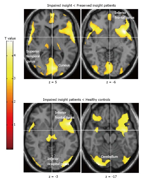
The three participant groups were comparable in age [F(2,57) = 0.34, P = 0.71] and the patient groups did not differ in age at illness onset [t(38) = 0.87, P = 0.39]. Insight(-) and Insight(+) patient groups also did not differ in symptoms on the Positive and Negative Syndromes scale (PANSS): Positive symptoms [t(38) = 0.58, P = 0.57], negative symptoms [t(38) = 0.61, P = 0.55], general psychopathology [t(38) = 1.30, P = 0.20] and total PANSS scores [t(38) = 0.21, P = 0.84]. The two patient groups, as expected, varied significantly in the level of BIS-assessed insight [t(38) = 12.11, P < 0.001]. MRI results revealed lower fronto-temporal, parahippocampal, occipital and cerebellar grey matter volumes in Insight(-) patients, relative to Insight(+) patients and healthy controls (for all clusters, family-wise error corrected P < 0.05). Insight(+) patient and healthy controls did not differ significantly (P > 0.20) from each other.
Our findings demonstrate a clear association between poor clinical insight and smaller fronto-temporal, occipital and cerebellar grey matter volumes in stable long-term schizophrenia patients.
定义精神分裂症患者中洞察力差(Insight(-))与洞察力正常(Insight(+))患者之间的区域性灰质异常,并与健康对照组进行比较。
对 40 例稳定期精神分裂症门诊患者(20 例 Insight(-)和 20 例 Insight(+))和 20 名健康对照者进行全脑磁共振成像(MRI)检查。采用 Birchwood 洞察力量表(BIS;一种自我报告测量工具)评估所有患者的洞察力。这两组患者在大多数临床和人口统计学参数上进行了预匹配,但通过设计,他们的 BIS 评分有明显的差异。SPM8 中采用的基于体素的形态计量学方法来检查整个大脑灰质体积的组间差异。
三组参与者的年龄相似[F(2,57) = 0.34,P = 0.71],且发病年龄在患者组间无差异[t(38) = 0.87,P = 0.39]。Insight(-)和 Insight(+)患者组在阳性和阴性综合征量表(PANSS)上的症状也没有差异:阳性症状[t(38) = 0.58,P = 0.57],阴性症状[t(38) = 0.61,P = 0.55],一般精神病学症状[t(38) = 1.30,P = 0.20]和总 PANSS 评分[t(38) = 0.21,P = 0.84]。正如预期的那样,这两组患者的 BIS 评估洞察力水平有显著差异[t(38) = 12.11,P < 0.001]。MRI 结果显示,与 Insight(+)患者和健康对照组相比,Insight(-)患者的额颞叶、海马旁回、枕叶和小脑灰质体积较低(所有聚类,经家族错误校正后 P < 0.05)。Insight(+)患者和健康对照组之间没有显著差异(P > 0.20)。
我们的研究结果表明,在稳定的长期精神分裂症患者中,临床洞察力差与额颞叶、枕叶和小脑灰质体积较小有明确的关联。