Li Kai-Ting, Chen Qing, Wang Da-Wu, Duan Qin-Qin, Tian Si, He Juan-Wen, Ou Yun-Sheng, Bai Ding-Qun
Department of Rehabilitation, The First Affiliated Hospital of Chongqing Medical University, Chongqing, China.
Department of Gastroenterology, Chinese Medicine Hospital of Longquan, Chengdu, China.
Cancer Med. 2016 Nov;5(11):3186-3193. doi: 10.1002/cam4.895. Epub 2016 Oct 3.
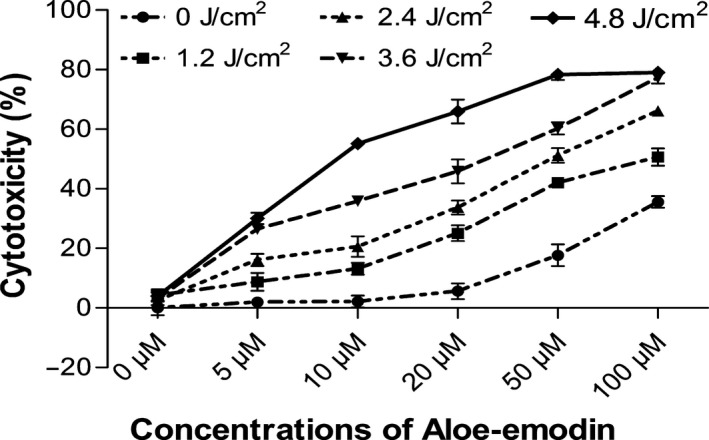
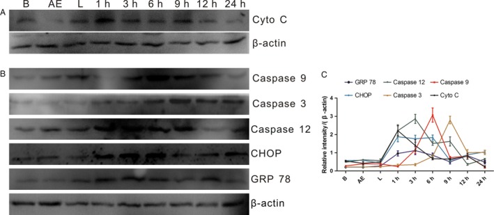
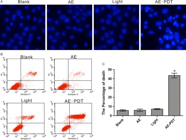
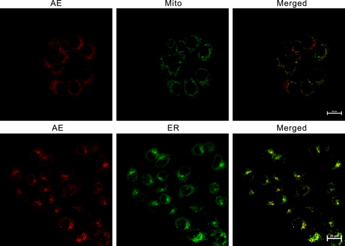
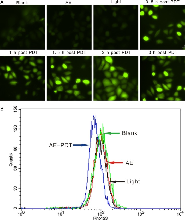
Photodynamic therapy (PDT) is a promising treatment in cancer therapy, with a photosensitizer activated by visible light. Aloe-emodin (AE) is a promising photosensitive agent. In this study, the photosensitizing effects and possible mechanisms of AE-PDT in MG63 cells were evaluated. The efficiency of AE-PDT was analyzed by MTT assay. The mode of cell death was investigated by Hoechst 33,342 staining and flow cytometer. The intracellular distribution of AE was detected with confocal microscopy. The formation of reactive oxygen species (ROS) was detected by DCFH-DA. The mitochondrial membrane potential (MMP) was measured by Rhodamine 123. The expression of proteins including cytochrome c, caspase-3, -9, and -12, CHOP and GRP78 was detected by western blot. Apoptosis is the primary mode of cell death in our study, which occurs in a manner of depending on AE concentration and irradiation dose. Confocal microscopy showed that AE was primarily localized on the mitochondria and endoplasmic reticulum (ER) of MG63 cells. AE-PDT resulted in rapid increases of intracellular ROS production, which reached a peak at 2 h, followed by declining of mitochondrial membrane potential, releasing of cytochrome c from mitochondria into the cytoplasm, and up-regulation of caspase-3, -9, and -12, CHOP and GRP78. These results suggest that death of MG63 cells induced by AE-PDT is triggered by ROS. Meanwhile, Mitochondria and ER serve as the subcellular targets, which are responsible for AE-PDT-induced death of MG63 cells.
光动力疗法(PDT)是癌症治疗中一种很有前景的治疗方法,通过可见光激活光敏剂。芦荟大黄素(AE)是一种很有前景的光敏剂。在本研究中,评估了AE-PDT对MG63细胞的光敏作用及其可能机制。通过MTT法分析AE-PDT的效率。通过Hoechst 33,342染色和流式细胞仪研究细胞死亡方式。用共聚焦显微镜检测AE的细胞内分布。用DCFH-DA检测活性氧(ROS)的形成。用罗丹明123测量线粒体膜电位(MMP)。通过蛋白质印迹法检测包括细胞色素c、半胱天冬酶-3、-9、-12、CHOP和GRP78等蛋白质的表达。在我们的研究中,凋亡是细胞死亡的主要方式,其发生方式取决于AE浓度和照射剂量。共聚焦显微镜显示,AE主要定位于MG63细胞的线粒体和内质网(ER)。AE-PDT导致细胞内ROS生成迅速增加,在2小时达到峰值,随后线粒体膜电位下降,细胞色素c从线粒体释放到细胞质中,以及半胱天冬酶-3、-9、-12、CHOP和GRP78上调。这些结果表明,AE-PDT诱导的MG63细胞死亡是由ROS触发的。同时,线粒体和内质网作为亚细胞靶点,负责AE-PDT诱导的MG63细胞死亡。