Olivier Juan-Gonzalo, García-Font Marc, Gonzalez-Sanchez Jose-Antonio, Roig-Cayon Miguel, Durán-Sindreu Fernando
DDS, PhD. Department of Restorative Dentistry and Endodontics, Universitat Internacional de Catalunya.
MD, PhD. Department of Restorative Dentistry and Endodontics, Universitat Internacional de Catalunya.
J Clin Exp Dent. 2016 Oct 1;8(4):e361-e367. doi: 10.4317/jced.52523. eCollection 2016 Oct.
The objective of the study was to evaluate and compare how apical enlargement with K3 and K3XF nickel-titanium (NiTi) rotary instruments reduces the root thickness in the danger zone and affects canal transportation and centering ability in mandibular molar mesial canals in a manikin extracted tooth model.
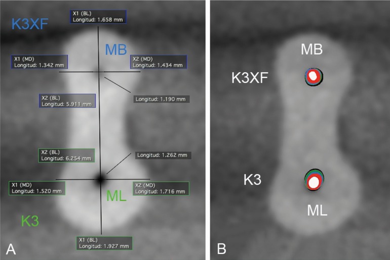
Seventy-two mesial root canals of first mandibular molars were instrumented. Initial and post-instrumentation Cone Beam Computed Tomography scans were performed after root canal preparation up to size 25, 30, 35 and 40 files. Canal transportation, canal centering and remaining root dentin thickness toward the danger zone were calculated in sections 1, 2 and 3 mm under the furcation level. Data were analyzed using non-parametric Kruskal-Wallis analysis of variance at a significance level of < 0.05.
K3 instruments removed more dentin toward the danger zone compared with K3XF instruments (< .05) and significant differences in dentin thickness were found when canal enlargement was performed to a #35-40 with both systems (< 0.05). No significant differences in canal transportation and centering ability were found between systems, except when canal enlargement was performed to a #40 ( = 0,0136). No differences were observed when comparing the number of uses in both systems (> 0.05).
Under the conditions of this study K3 removed a significant amount of dentin at the furcation level compared with the R-Phase K3XF rotary system in curved root canals. Enlargement to a 35-40/04 file removed significantly more dentin with both systems. K3, K3XF, R-phase, center ability, canal transportation, dentin thickness, increased apical enlargement, danger zone, dentin thickness.
本研究的目的是评估和比较使用K3和K3XF镍钛(NiTi)旋转器械进行根尖扩大时,在人体模拟离体牙模型中,下颌磨牙近中根管危险区的牙根厚度如何减小,以及对根管偏移和根管居中能力的影响。
对72颗下颌第一磨牙的近中根管进行预备。在根管预备至25、30、35和40号锉后,进行初始和预备后锥形束计算机断层扫描。在根分叉水平以下1、2和3mm处的截面计算根管偏移、根管居中情况以及朝向危险区的剩余牙根牙本质厚度。使用非参数Kruskal-Wallis方差分析对数据进行分析,显著性水平<0.05。
与K3XF器械相比,K3器械向危险区去除了更多牙本质(<0.05),并且当两种系统均将根管扩大至35 - 40号时,牙本质厚度存在显著差异(<0.05)。除了将根管扩大至40号时(P = 0.0136),两种系统在根管偏移和根管居中能力方面未发现显著差异。比较两种系统的使用次数时未观察到差异(>0.05)。
在本研究条件下,与R相K3XF旋转系统相比,K3在弯曲根管的根分叉水平去除了大量牙本质。两种系统将根管扩大至35 - 40/04号锉时,去除的牙本质显著更多。K3、K3XF、R相、居中能力、根管偏移、牙本质厚度、根尖扩大增加、危险区、牙本质厚度