• 文献检索
  • 文档翻译
  • 深度研究
  • 学术资讯
  • Suppr Zotero 插件Zotero 插件
  • 邀请有礼
  • 套餐&价格
  • 历史记录
应用&插件
Suppr Zotero 插件Zotero 插件浏览器插件Mac 客户端Windows 客户端微信小程序
定价
高级版会员购买积分包购买API积分包
服务
文献检索文档翻译深度研究API 文档MCP 服务
关于我们
关于 Suppr公司介绍联系我们用户协议隐私条款
关注我们

Suppr 超能文献

核心技术专利:CN118964589B侵权必究
粤ICP备2023148730 号-1Suppr @ 2026

文献检索

告别复杂PubMed语法,用中文像聊天一样搜索,搜遍4000万医学文献。AI智能推荐,让科研检索更轻松。

立即免费搜索

文件翻译

保留排版,准确专业,支持PDF/Word/PPT等文件格式,支持 12+语言互译。

免费翻译文档

深度研究

AI帮你快速写综述,25分钟生成高质量综述,智能提取关键信息,辅助科研写作。

立即免费体验

扩散加权成像:技术与应用

Diffusion weighted imaging: Technique and applications.

作者信息

Baliyan Vinit, Das Chandan J, Sharma Raju, Gupta Arun Kumar

机构信息

Vinit Baliyan, Chandan J Das, Raju Sharma, Arun Kumar Gupta, Department of Radiology, All India Institute of Medical Sciences, New Delhi 110029, India.

出版信息

World J Radiol. 2016 Sep 28;8(9):785-798. doi: 10.4329/wjr.v8.i9.785.

DOI:10.4329/wjr.v8.i9.785
PMID:27721941
原文链接:https://pmc.ncbi.nlm.nih.gov/articles/PMC5039674/
Abstract

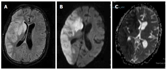
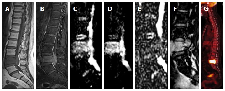
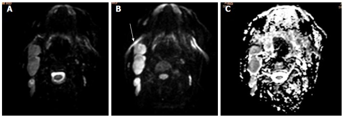
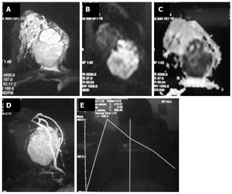
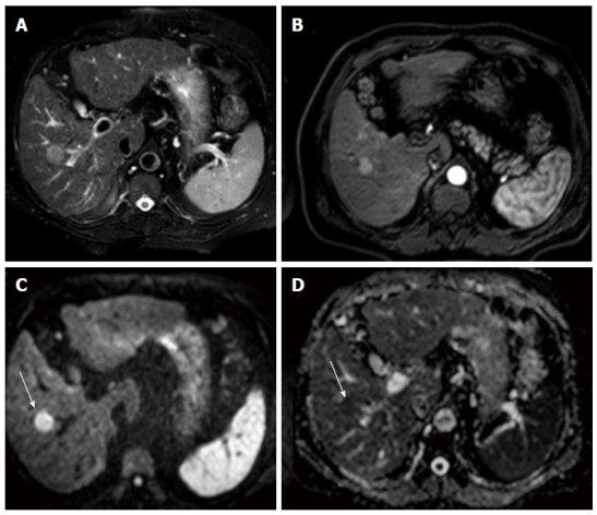
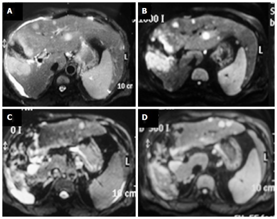
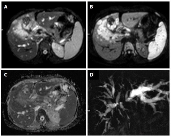
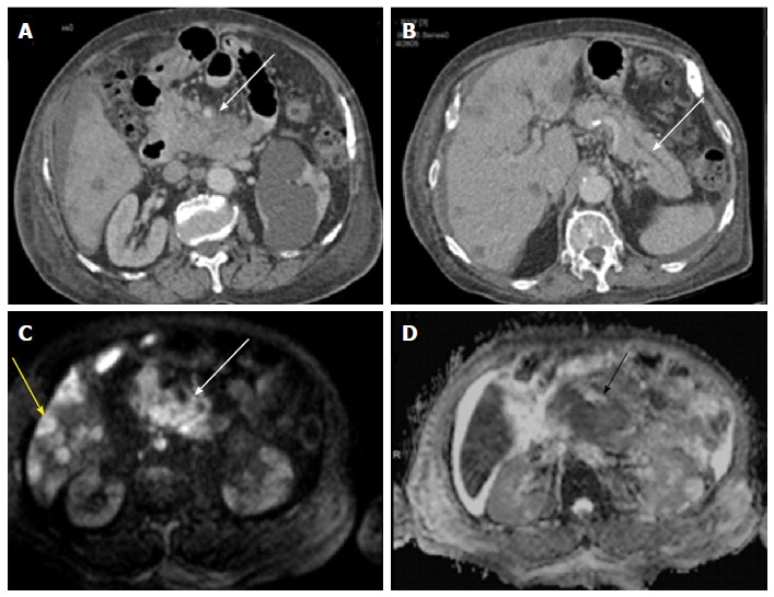
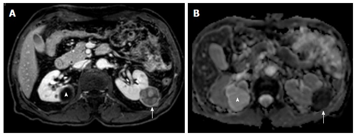
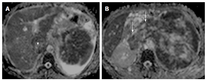
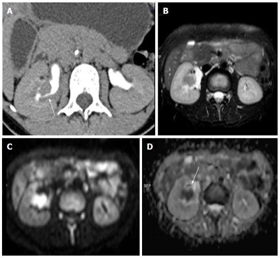
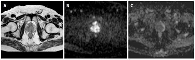
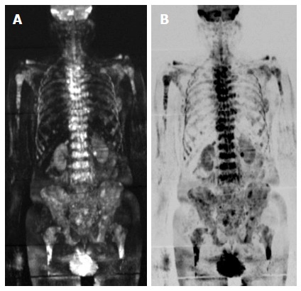
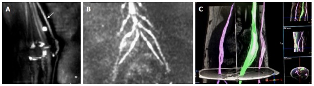
Diffusion weighted imaging (DWI) is a method of signal contrast generation based on the differences in Brownian motion. DWI is a method to evaluate the molecular function and micro-architecture of the human body. DWI signal contrast can be quantified by apparent diffusion coefficient maps and it acts as a tool for treatment response evaluation and assessment of disease progression. Ability to detect and quantify the anisotropy of diffusion leads to a new paradigm called diffusion tensor imaging (DTI). DTI is a tool for assessment of the organs with highly organised fibre structure. DWI forms an integral part of modern state-of-art magnetic resonance imaging and is indispensable in neuroimaging and oncology. DWI is a field that has been undergoing rapid technical evolution and its applications are increasing every day. This review article provides insights in to the evolution of DWI as a new imaging paradigm and provides a summary of current role of DWI in various disease processes.

摘要

扩散加权成像(DWI)是一种基于布朗运动差异产生信号对比的方法。DWI是一种评估人体分子功能和微观结构的方法。DWI信号对比可通过表观扩散系数图进行量化,它是用于评估治疗反应和疾病进展的工具。检测和量化扩散各向异性的能力导致了一种称为扩散张量成像(DTI)的新范式。DTI是一种用于评估具有高度有组织纤维结构器官的工具。DWI是现代先进磁共振成像不可或缺的一部分,在神经成像和肿瘤学中必不可少。DWI是一个技术正在迅速发展的领域,其应用日益增多。这篇综述文章深入探讨了DWI作为一种新成像范式的发展历程,并总结了DWI在各种疾病过程中的当前作用。

https://cdn.ncbi.nlm.nih.gov/pmc/blobs/a94d/5039674/281debef4752/WJR-8-785-g015.jpg
https://cdn.ncbi.nlm.nih.gov/pmc/blobs/a94d/5039674/098da47ea046/WJR-8-785-g001.jpg
https://cdn.ncbi.nlm.nih.gov/pmc/blobs/a94d/5039674/e808cb5df00c/WJR-8-785-g002.jpg
https://cdn.ncbi.nlm.nih.gov/pmc/blobs/a94d/5039674/e6a9975230b0/WJR-8-785-g003.jpg
https://cdn.ncbi.nlm.nih.gov/pmc/blobs/a94d/5039674/0a92f0341bd7/WJR-8-785-g004.jpg
https://cdn.ncbi.nlm.nih.gov/pmc/blobs/a94d/5039674/a98a93678ccb/WJR-8-785-g005.jpg
https://cdn.ncbi.nlm.nih.gov/pmc/blobs/a94d/5039674/d809943ce221/WJR-8-785-g006.jpg
https://cdn.ncbi.nlm.nih.gov/pmc/blobs/a94d/5039674/86f2441afd9f/WJR-8-785-g007.jpg
https://cdn.ncbi.nlm.nih.gov/pmc/blobs/a94d/5039674/feb850746503/WJR-8-785-g008.jpg
https://cdn.ncbi.nlm.nih.gov/pmc/blobs/a94d/5039674/445d0bf18014/WJR-8-785-g009.jpg
https://cdn.ncbi.nlm.nih.gov/pmc/blobs/a94d/5039674/d845316a9270/WJR-8-785-g010.jpg
https://cdn.ncbi.nlm.nih.gov/pmc/blobs/a94d/5039674/175adea32fa8/WJR-8-785-g011.jpg
https://cdn.ncbi.nlm.nih.gov/pmc/blobs/a94d/5039674/8ffb18657914/WJR-8-785-g012.jpg
https://cdn.ncbi.nlm.nih.gov/pmc/blobs/a94d/5039674/35bb52fc53ab/WJR-8-785-g013.jpg
https://cdn.ncbi.nlm.nih.gov/pmc/blobs/a94d/5039674/9c8cc5a94825/WJR-8-785-g014.jpg
https://cdn.ncbi.nlm.nih.gov/pmc/blobs/a94d/5039674/281debef4752/WJR-8-785-g015.jpg

相似文献

1
Diffusion weighted imaging: Technique and applications.扩散加权成像:技术与应用
World J Radiol. 2016 Sep 28;8(9):785-798. doi: 10.4329/wjr.v8.i9.785.
2
[Basic principles and technique of diffusion-weighted imaging and diffusion tensor imaging].[扩散加权成像与扩散张量成像的基本原理及技术]
Radiologe. 2015 Sep;55(9):762-70. doi: 10.1007/s00117-015-0004-7.
3
Diffusion-weighted and diffusion-tensor imaging of normal and diseased uterus.正常及病变子宫的扩散加权成像和扩散张量成像
World J Radiol. 2015 Jul 28;7(7):149-56. doi: 10.4329/wjr.v7.i7.149.
4
Comparison of the diagnostic performances of diffusion parameters in diffusion weighted imaging and diffusion tensor imaging of breast lesions.扩散加权成像和扩散张量成像中扩散参数对乳腺病变诊断性能的比较。
Eur J Radiol. 2013 Dec;82(12):e801-6. doi: 10.1016/j.ejrad.2013.09.001. Epub 2013 Sep 13.
5
Differential diagnosis of intracranial ring enhancing cystic mass lesions--role of diffusion-weighted imaging (DWI) and diffusion-tensor imaging (DTI).颅内环形强化囊性肿块病变的鉴别诊断——弥散加权成像(DWI)和弥散张量成像(DTI)的作用
Clin Neurol Neurosurg. 2010 Apr;112(3):218-25. doi: 10.1016/j.clineuro.2009.11.016. Epub 2010 Jan 6.
6
Diffusion-weighted imaging and diffusion tensor imaging of the heart : major developments.心脏的扩散加权成像和扩散张量成像:主要进展
Postepy Kardiol Interwencyjnej. 2022 Dec;18(4):350-359. doi: 10.5114/aic.2022.121345. Epub 2022 Nov 19.
7
Diffusion-weighted imaging in urinary tract lesions.泌尿系统病变的扩散加权成像
Clin Radiol. 2014 Aug;69(8):773-82. doi: 10.1016/j.crad.2014.01.011. Epub 2014 Feb 26.
8
Technical aspects of MR diffusion imaging of the body.身体磁共振弥散成像技术方面。
Eur J Radiol. 2010 Dec;76(3):314-22. doi: 10.1016/j.ejrad.2010.02.018. Epub 2010 Mar 29.
9
Utility of the Diffusion Weighted Sequence in Gynecological Imaging: Review Article.扩散加权序列在妇科成像中的应用:综述文章。
Cancers (Basel). 2022 Sep 15;14(18):4468. doi: 10.3390/cancers14184468.
10
Assessment of brain maturation in the preterm infants using diffusion tensor imaging (DTI) and enhanced T2 star weighted angiography (ESWAN).应用弥散张量成像(DTI)和增强 T2*星加权血管造影(ESWAN)评估早产儿脑成熟度。
Eur J Radiol. 2013 Sep;82(9):e476-83. doi: 10.1016/j.ejrad.2013.04.003. Epub 2013 Apr 29.

引用本文的文献

1
Exploring mean kurtosis in MR diffusion kurtosis imaging for early detection of lumbar spine degeneration: a systematic review.探索磁共振扩散峰度成像中的平均峰度用于早期检测腰椎退变:一项系统综述
F1000Res. 2025 Apr 15;14:440. doi: 10.12688/f1000research.163638.1. eCollection 2025.
2
Diffusion weighted imaging in musculoskeletal system: where are we now?肌肉骨骼系统中的扩散加权成像:我们目前处于什么阶段?
BJR Open. 2025 Jul 22;7(1):tzaf019. doi: 10.1093/bjro/tzaf019. eCollection 2025 Jan.
3
Adaptive Radiation Therapy for Head and Neck Cancer.

本文引用的文献

1
Diffusion weighted imaging in gynecological malignancies - present and future.妇科恶性肿瘤中的弥散加权成像——现状与未来
World J Radiol. 2016 Mar 28;8(3):288-97. doi: 10.4329/wjr.v8.i3.288.
2
Magnetic resonance imaging for N staging in non-small cell lung cancer: A systematic review and meta-analysis.磁共振成像在非小细胞肺癌 N 分期中的应用:系统评价和荟萃分析。
Thorac Cancer. 2015 Mar;6(2):123-32. doi: 10.1111/1759-7714.12203. Epub 2015 Jan 8.
3
Diffusion-weighted and PET/MR Imaging after Radiation Therapy for Malignant Head and Neck Tumors.
头颈部癌的自适应放射治疗
ArXiv. 2025 Aug 1:arXiv:2508.00651v1.
4
Magnetic Resonance Imaging in the Evaluation of the Stress System in Acute and Chronic Cardiac Disease.磁共振成像在急慢性心脏疾病应激系统评估中的应用
Diagnostics (Basel). 2025 Jul 4;15(13):1712. doi: 10.3390/diagnostics15131712.
5
Dynamic changes in BDNF, VEGF, and GDNF after transplanting human protein-based scaffolds with Wharton's Jelly MSCs in a rat brain injury model.在大鼠脑损伤模型中移植含华通氏胶间充质干细胞的基于人类蛋白质的支架后,脑源性神经营养因子(BDNF)、血管内皮生长因子(VEGF)和胶质细胞源性神经营养因子(GDNF)的动态变化。
Sci Rep. 2025 Jul 2;15(1):22625. doi: 10.1038/s41598-025-04269-w.
6
Imaging markers of neuroinflammation in aging and Alzheimer's disease and related dementia: A comprehensive review.衰老、阿尔茨海默病及相关痴呆症中神经炎症的影像学标志物:综述
Neurosci Biobehav Rev. 2025 Jun 25;176:106270. doi: 10.1016/j.neubiorev.2025.106270.
7
The Acute Effects of Non-concussive Head Impacts on Brain Microstructure, Chemistry and Function in Male Soccer Players: A Pilot Randomised Controlled Trial.非震荡性头部撞击对男性足球运动员脑微观结构、化学组成和功能的急性影响:一项初步随机对照试验
Sports Med Open. 2025 Jun 18;11(1):77. doi: 10.1186/s40798-025-00867-0.
8
Multiparametric MRI in Diagnosis of Parotid Gland Tumor: An Observational Study in 3-T MRI.多参数磁共振成像在腮腺肿瘤诊断中的应用:一项3-T磁共振成像的观察性研究
Indian J Radiol Imaging. 2024 Dec 17;35(3):402-410. doi: 10.1055/s-0044-1800861. eCollection 2025 Jul.
9
The Evolving Landscape of Ovarian Cancer: Innovations in Biotechnology and Artificial Intelligence- Based Screening and Treatment.卵巢癌的发展态势:基于生物技术和人工智能的筛查与治疗创新
Curr Treat Options Oncol. 2025 Jun 9. doi: 10.1007/s11864-025-01331-7.
10
Differential tractography: an imaging marker for tissue degeneration in neurodegenerative diseases.差异纤维束成像:神经退行性疾病中组织退化的一种影像学标志物。
Brain Commun. 2025 May 24;7(3):fcaf198. doi: 10.1093/braincomms/fcaf198. eCollection 2025.
恶性头颈部肿瘤放疗后的扩散加权成像及PET/MR成像
Radiographics. 2015 Sep-Oct;35(5):1502-27. doi: 10.1148/rg.2015140029. Epub 2015 Aug 7.
4
Diffusion-weighted MR imaging using FASE sequence for 3T MR system: Preliminary comparison of capability for N-stage assessment by means of diffusion-weighted MR imaging using EPI sequence, STIR FASE imaging and FDG PET/CT for non-small cell lung cancer patients.使用3T MR系统的快速自旋回波(FASE)序列进行扩散加权磁共振成像:通过使用回波平面成像(EPI)序列的扩散加权磁共振成像、短TI反转恢复快速自旋回波(STIR FASE)成像和氟代脱氧葡萄糖正电子发射断层显像/计算机断层扫描(FDG PET/CT)对非小细胞肺癌患者进行N分期评估能力的初步比较。
Eur J Radiol. 2015 Nov;84(11):2321-31. doi: 10.1016/j.ejrad.2015.07.019. Epub 2015 Jul 26.
5
Peripheral Nerve Diffusion Tensor Imaging: Assessment of Axon and Myelin Sheath Integrity.周围神经扩散张量成像:轴突和髓鞘完整性评估
PLoS One. 2015 Jun 26;10(6):e0130833. doi: 10.1371/journal.pone.0130833. eCollection 2015.
6
Prognostication and response assessment in liver and pancreatic tumors: The new imaging.肝脏和胰腺肿瘤的预后评估与疗效评估:新型影像学检查
World J Gastroenterol. 2015 Jun 14;21(22):6794-808. doi: 10.3748/wjg.v21.i22.6794.
7
Differential diagnosis of pancreatic cancer by single-shot echo-planar imaging diffusion-weighted imaging.单次激发回波平面成像扩散加权成像对胰腺癌的鉴别诊断
World J Gastroenterol. 2015 May 28;21(20):6374-80. doi: 10.3748/wjg.v21.i20.6374.
8
Diffusion weighted magnetic resonance imaging and its recent trend-a survey.磁共振扩散加权成像及其近期趋势——一项综述
Quant Imaging Med Surg. 2015 Jun;5(3):407-22. doi: 10.3978/j.issn.2223-4292.2015.03.01.
9
Apparent diffusion coefficient values of diffusion-weighted imaging for distinguishing focal pulmonary lesions and characterizing the subtype of lung cancer: a meta-analysis.弥散加权成像表观扩散系数值鉴别肺部局灶性病变及肺癌亚型的特征:荟萃分析。
Eur Radiol. 2016 Feb;26(2):556-66. doi: 10.1007/s00330-015-3840-y. Epub 2015 May 24.
10
Diffusion-weighted MRI in pediatric inflammatory bowel disease.磁共振弥散加权成像在儿科炎症性肠病中的应用。
AJR Am J Roentgenol. 2015 Jun;204(6):1269-77. doi: 10.2214/AJR.14.13359.