• 文献检索
  • 文档翻译
  • 深度研究
  • 学术资讯
  • Suppr Zotero 插件Zotero 插件
  • 邀请有礼
  • 套餐&价格
  • 历史记录
应用&插件
Suppr Zotero 插件Zotero 插件浏览器插件Mac 客户端Windows 客户端微信小程序
定价
高级版会员购买积分包购买API积分包
服务
文献检索文档翻译深度研究API 文档MCP 服务
关于我们
关于 Suppr公司介绍联系我们用户协议隐私条款
关注我们

Suppr 超能文献

核心技术专利:CN118964589B侵权必究
粤ICP备2023148730 号-1Suppr @ 2026

文献检索

告别复杂PubMed语法,用中文像聊天一样搜索,搜遍4000万医学文献。AI智能推荐,让科研检索更轻松。

立即免费搜索

文件翻译

保留排版,准确专业,支持PDF/Word/PPT等文件格式,支持 12+语言互译。

免费翻译文档

深度研究

AI帮你快速写综述,25分钟生成高质量综述,智能提取关键信息,辅助科研写作。

立即免费体验

脊柱和骶髂关节炎症性与退行性MRI表现亚组的识别:对1037例持续性下腰痛患者的潜在类别分析

Identification of subgroups of inflammatory and degenerative MRI findings in the spine and sacroiliac joints: a latent class analysis of 1037 patients with persistent low back pain.

作者信息

Arnbak Bodil, Jensen Rikke Krüger, Manniche Claus, Hendricks Oliver, Kent Peter, Jurik Anne Grethe, Jensen Tue Secher

机构信息

Research Department, Spine Centre of Southern Denmark, Hospital Lillebaelt, Oestre Hougvej 55, Middelfart, 5500, Denmark.

Institute of Regional Health Research, University of Southern Denmark, Winsloewparken 19-3, Odense C, 5000, Denmark.

出版信息

Arthritis Res Ther. 2016 Oct 13;18(1):237. doi: 10.1186/s13075-016-1131-x.

DOI:10.1186/s13075-016-1131-x
PMID:27733191
原文链接:https://pmc.ncbi.nlm.nih.gov/articles/PMC5062874/
Abstract

BACKGROUND

The aim of this study was to investigate subgroups of magnetic resonance imaging (MRI) findings for the spine and sacroiliac joints (SIJs) using latent class analysis (LCA), and to investigate whether these subgroups differ in their demographic and clinical characteristics.

METHODS

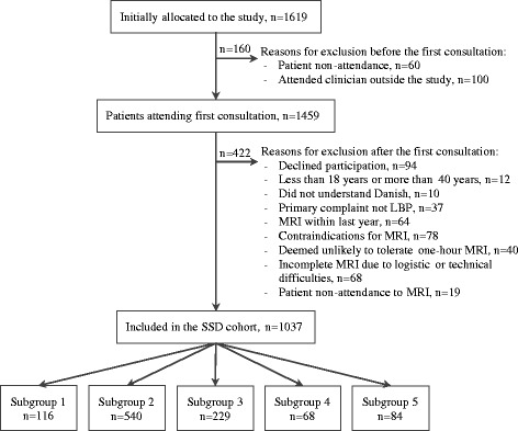
The sample included 1037 patients aged 18-40 years with persistent low back pain (LBP). LCA was applied to MRI findings of the spine and SIJs. The resulting subgroups were tested for differences in self-reported demographic and clinical characteristics.

RESULTS

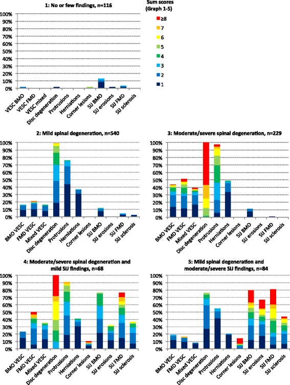
A five-class model was identified: Subgroup 1, 'No or few findings' (n = 116); Subgroup 2, 'Mild spinal degeneration' (n = 540); Subgroup 3, 'Moderate to severe spinal degeneration' (n = 229); Subgroup 4, 'Moderate to severe spinal degeneration with mild SIJ findings' (n = 68); and Subgroup 5, 'Mild spinal degeneration with moderate to severe SIJ findings' (n = 84). The two SIJ subgroups (Subgroups 4 and 5) had a higher median activity limitation score (Roland Morris Disability Questionnaire calculated as a proportional score: 65 (IQR 48-78)/65 (48-78)) compared with Subgroups 1-3 (48 (35-74)/57 (39-74)/57 (39-74)), a higher prevalence of women (68 % (95 % CI 56-79)/68 % (58-78)) compared with Subgroups 2 and 3 (51 % (47-55)/40 % (33-46)), a higher prevalence of being overweight (67 % (95 % CI 55-79)/53 % (41-65)) compared with Subgroup 1 (36 % (26-46)) and a higher prevalence of previous LBP episodes (yes/no: 81 % (95 % CI 71-91)/79 % (70-89)) compared with Subgroup 1 (58 % (48-67)). Subgroup 5 was younger than Subgroup 4 (median age 29 years (IQR 25-33) versus 34 years (30-37)) and had a higher prevalence of HLA-B27 (40 % (95 % CI 29-50)) compared with the other subgroups (Subgroups 1-4: 12 % (6-18)/7 % (5-10)/6 % (3-9)/12 % (4-20)). Across the subgroups with predominantly spinal findings (Subgroups 1-3), median age, prevalence of men, being overweight and previous LBP episodes were statistically significantly lower in Subgroup 1, higher in Subgroup 2 and highest in Subgroup 3.

CONCLUSIONS

Five distinct subgroups of MRI findings in the spine and SIJs were identified. The results indicate that SIJ MRI findings not only can be seen as a part of the spondyloarthritis disease entity, but also are associated with age, gender and being overweight. Furthermore, the results indicate that LBP patients with SIJ MRI findings are more disabled compared with patients without SIJ MRI findings, and that moderate to severe spinal degeneration and/or SIJ MRI findings may be associated with recurrent pain.

摘要

背景

本研究旨在利用潜在类别分析(LCA)探究脊柱和骶髂关节(SIJ)磁共振成像(MRI)结果的亚组,并研究这些亚组在人口统计学和临床特征方面是否存在差异。

方法

样本包括1037名年龄在18至40岁之间的持续性下腰痛(LBP)患者。将LCA应用于脊柱和SIJ的MRI结果。对所得亚组的自我报告人口统计学和临床特征差异进行测试。

结果

确定了一个五类模型:亚组1,“无或少量发现”(n = 116);亚组2,“轻度脊柱退变”(n = 540);亚组3,“中度至重度脊柱退变”(n = 229);亚组4,“中度至重度脊柱退变伴轻度SIJ发现”(n = 68);亚组5,“轻度脊柱退变伴中度至重度SIJ发现”(n = 84)。与亚组1至3(48(35 - 74)/57(39 - 74)/57(39 - 74))相比,两个SIJ亚组(亚组4和5)的中位活动受限评分更高(罗兰·莫里斯残疾问卷以比例评分计算:65(四分位间距48 - 78)/65(48 - 78)),女性患病率更高(68%(95%置信区间56 - 79)/68%(58 - 78)),相比之下亚组2和3为51%(47 - 55)/40%(33 - 46)),超重患病率更高(67%(95%置信区间55 - 79)/53%(41 - 65)),相比之下亚组1为36%(26 - 46)),既往LBP发作患病率更高(是/否:81%(95%置信区间71 - 91)/79%(70 - 89)),相比之下亚组1为58%(48 - 67))。亚组5比亚组4年轻(中位年龄29岁(四分位间距25 - 33)对34岁(30 - 37)),与其他亚组(亚组1至4:12%(6 - 18)/7%(5 - 10)/6%(3 - 9)/12%(4 - 20))相比,HLA - B27患病率更高(40%(95%置信区间29 - 50))。在主要为脊柱发现的亚组(亚组1至3)中,亚组1的中位年龄、男性患病率、超重和既往LBP发作在统计学上显著更低,亚组2更高,亚组3最高。

结论

确定了脊柱和SIJ的MRI结果的五个不同亚组。结果表明,SIJ的MRI发现不仅可被视为脊柱关节炎疾病实体的一部分,还与年龄、性别和超重有关。此外,结果表明与无SIJ的MRI发现的患者相比,有SIJ的MRI发现的LBP患者残疾程度更高,并且中度至重度脊柱退变和/或SIJ的MRI发现可能与复发性疼痛有关。

https://cdn.ncbi.nlm.nih.gov/pmc/blobs/c7df/5062874/2dcdcd1a3808/13075_2016_1131_Fig2_HTML.jpg
https://cdn.ncbi.nlm.nih.gov/pmc/blobs/c7df/5062874/af017c8ae672/13075_2016_1131_Fig1_HTML.jpg
https://cdn.ncbi.nlm.nih.gov/pmc/blobs/c7df/5062874/2dcdcd1a3808/13075_2016_1131_Fig2_HTML.jpg
https://cdn.ncbi.nlm.nih.gov/pmc/blobs/c7df/5062874/af017c8ae672/13075_2016_1131_Fig1_HTML.jpg
https://cdn.ncbi.nlm.nih.gov/pmc/blobs/c7df/5062874/2dcdcd1a3808/13075_2016_1131_Fig2_HTML.jpg

相似文献

1
Identification of subgroups of inflammatory and degenerative MRI findings in the spine and sacroiliac joints: a latent class analysis of 1037 patients with persistent low back pain.脊柱和骶髂关节炎症性与退行性MRI表现亚组的识别:对1037例持续性下腰痛患者的潜在类别分析
Arthritis Res Ther. 2016 Oct 13;18(1):237. doi: 10.1186/s13075-016-1131-x.
2
Prevalence of degenerative and spondyloarthritis-related magnetic resonance imaging findings in the spine and sacroiliac joints in patients with persistent low back pain.持续性下腰痛患者脊柱和骶髂关节中与退行性和脊柱关节炎相关的磁共振成像表现的患病率。
Eur Radiol. 2016 Apr;26(4):1191-203. doi: 10.1007/s00330-015-3903-0. Epub 2015 Jul 22.
3
Spine and Sacroiliac Joints Lesions on Magnetic Resonance Imaging in Early Axial-Spondyloarthritis During 24-Months Follow-Up (Italian Arm of SPACE Study).脊柱和骶髂关节磁共振成像在 24 个月随访中早期中轴型脊柱关节炎的病变(SPACE 研究意大利分支)。
Front Immunol. 2020 May 15;11:936. doi: 10.3389/fimmu.2020.00936. eCollection 2020.
4
Prevalence and awareness of sacroiliac joint alterations on lumbar spine CT in low back pain patients younger than 40 years.40岁以下腰痛患者腰椎CT上骶髂关节改变的患病率及知晓率
Acta Radiol. 2017 Apr;58(4):449-455. doi: 10.1177/0284185116656490. Epub 2016 Jul 28.
5
Does spinal MRI add incremental diagnostic value to MRI of the sacroiliac joints alone in patients with non-radiographic axial spondyloarthritis?对于非放射性轴性脊柱关节炎患者,脊柱 MRI 是否比单独的骶髂关节 MRI 具有更多的附加诊断价值?
Ann Rheum Dis. 2015 Jun;74(6):985-92. doi: 10.1136/annrheumdis-2013-203887. Epub 2014 Jan 22.
6
Spine and sacroiliac joints on magnetic resonance imaging in patients with early axial spondyloarthritis: prevalence of lesions and association with clinical and disease activity indices from the Italian group of the SPACE study.早期轴性脊柱关节炎患者脊柱和骶髂关节的磁共振成像:SPACE研究意大利组中病变的患病率及其与临床和疾病活动指数的关联
Reumatismo. 2016 Sep 9;68(2):72-82. doi: 10.4081/reumatismo.2016.885.
7
Fat Metaplasia on Sacroiliac Joint Magnetic Resonance Imaging at Baseline Is Associated with Spinal Radiographic Progression in Patients with Axial Spondyloarthritis.基线时骶髂关节磁共振成像上的脂肪化生与轴性脊柱关节炎患者的脊柱放射学进展相关。
PLoS One. 2015 Aug 13;10(8):e0135206. doi: 10.1371/journal.pone.0135206. eCollection 2015.
8
Associations Between Spondyloarthritis Features and Magnetic Resonance Imaging Findings: A Cross-Sectional Analysis of 1,020 Patients With Persistent Low Back Pain.脊柱关节炎特征与磁共振成像结果之间的关联:1020 例持续性腰痛患者的横断面分析。
Arthritis Rheumatol. 2016 Apr;68(4):892-900. doi: 10.1002/art.39551.
9
Structural progression rate decreases over time on serial radiography and magnetic resonance imaging of sacroiliac joints and spine in a five-year follow-up study of patients with ankylosing spondylitis treated with tumour necrosis factor inhibitor.在一项为期五年的强直性脊柱炎患者肿瘤坏死因子抑制剂治疗的随访研究中,通过骶髂关节和脊柱的连续影像学(X 线和磁共振)发现,结构进展率随时间推移而降低。
Scand J Rheumatol. 2019 May;48(3):185-197. doi: 10.1080/03009742.2018.1506822. Epub 2018 Nov 13.
10
Does the site of magnetic resonance imaging abnormalities match the site of recent-onset inflammatory back pain? The DESIR cohort.磁共振成像异常部位与近期发作的炎症性背痛部位是否一致?DESIR 队列研究。
Ann Rheum Dis. 2013 Jun;72(6):979-85. doi: 10.1136/annrheumdis-2012-201427. Epub 2012 Aug 14.

引用本文的文献

1
Associations Between Sum Scores or Combinations of MRI Findings in the Lumbar Spine and Low Back Pain-Related Outcomes: A Systematic Review.腰椎MRI检查结果总和评分或组合与下腰痛相关结局之间的关联:一项系统评价
Eur J Pain. 2025 Aug;29(7):e70076. doi: 10.1002/ejp.70076.
2
Where are the chiropractic clinical outcomes registries? A scoping review.脊椎按摩疗法临床结果登记处在哪里?一项范围综述。
Chiropr Man Therap. 2025 May 25;33(1):22. doi: 10.1186/s12998-025-00583-2.
3
Human Disc Degeneration Is Accompanied by a Loss of Anterior Annulus Fibrosus Interlamellar Matrix Integrity as Assessed by Peel Tests.

本文引用的文献

1
Imaging in Spondyloarthritis: Controversies in Recognition of Early Disease.脊柱关节炎的影像学检查:早期疾病识别中的争议
Curr Rheumatol Rep. 2016 Sep;18(9):58. doi: 10.1007/s11926-016-0607-7.
2
Trends in adult body-mass index in 200 countries from 1975 to 2014: a pooled analysis of 1698 population-based measurement studies with 19·2 million participants.1975年至2014年200个国家成人身体质量指数的趋势:对1698项基于人群测量研究的汇总分析,涉及1920万参与者。
Lancet. 2016 Apr 2;387(10026):1377-1396. doi: 10.1016/S0140-6736(16)30054-X.
3
Associations Between Spondyloarthritis Features and Magnetic Resonance Imaging Findings: A Cross-Sectional Analysis of 1,020 Patients With Persistent Low Back Pain.
通过剥离试验评估发现,人类椎间盘退变伴随着前侧纤维环板层间基质完整性的丧失。
JOR Spine. 2025 May 14;8(2):e70067. doi: 10.1002/jsp2.70067. eCollection 2025 Jun.
4
Collagen integrity of the annulus fibrosus in degenerative disc disease individuals quantified with collagen hybridizing peptide.用胶原杂交肽定量分析退变椎间盘个体纤维环的胶原完整性。
JOR Spine. 2024 Jul 31;7(3):e1359. doi: 10.1002/jsp2.1359. eCollection 2024 Sep.
5
Mast Cells and Their Related Gene HK-1 are Closely Associated with Discogenic Low Back Pain: A Bioinformatics and Clinical Sample Study.肥大细胞及其相关基因HK-1与椎间盘源性下腰痛密切相关:一项生物信息学与临床样本研究
J Pain Res. 2024 Apr 8;17:1401-1412. doi: 10.2147/JPR.S454785. eCollection 2024.
6
Macrophages and Intervertebral Disc Degeneration.巨噬细胞与椎间盘退变。
Int J Mol Sci. 2023 Jan 10;24(2):1367. doi: 10.3390/ijms24021367.
7
Cultural adaptation and psychometric assessment of the Persian version of the lumbar spine instability questionnaire.腰椎不稳定问卷波斯语版本的文化调适与心理测量评估
BMC Sports Sci Med Rehabil. 2022 May 19;14(1):90. doi: 10.1186/s13102-022-00486-2.
8
Intervertebral Disc Degeneration: Biomaterials and Tissue Engineering Strategies toward Precision Medicine.椎间盘退变性疾病:精准医学的生物材料和组织工程策略。
Adv Healthc Mater. 2022 Jul;11(13):e2102530. doi: 10.1002/adhm.202102530. Epub 2022 May 4.
9
Painful intervertebral disc degeneration and inflammation: from laboratory evidence to clinical interventions.疼痛性椎间盘退变与炎症:从实验室证据到临床干预
Bone Res. 2021 Jan 29;9(1):7. doi: 10.1038/s41413-020-00125-x.
10
Improving the staging of neck injuries using a new index, the Neck Functional Holistic Analysis Score: Clustering approach to determine degrees of impairment.采用新的颈部功能整体分析评分(Neck Functional Holistic Analysis Score)指数提高颈部损伤分期:聚类方法确定损伤程度。
PLoS One. 2020 Sep 9;15(9):e0238424. doi: 10.1371/journal.pone.0238424. eCollection 2020.
脊柱关节炎特征与磁共振成像结果之间的关联:1020 例持续性腰痛患者的横断面分析。
Arthritis Rheumatol. 2016 Apr;68(4):892-900. doi: 10.1002/art.39551.
4
Modic changes of the lumbar spine: prevalence, risk factors, and association with disc degeneration and low back pain in a large-scale population-based cohort.腰椎的Modic改变:大规模人群队列中的患病率、危险因素及其与椎间盘退变和腰痛的关联
Spine J. 2016 Jan 1;16(1):32-41. doi: 10.1016/j.spinee.2015.09.060. Epub 2015 Oct 8.
5
SpineData - a Danish clinical registry of people with chronic back pain.脊柱数据——丹麦慢性背痛患者临床登记处。
Clin Epidemiol. 2015 Aug 13;7:369-80. doi: 10.2147/CLEP.S83830. eCollection 2015.
6
Prevalence of degenerative and spondyloarthritis-related magnetic resonance imaging findings in the spine and sacroiliac joints in patients with persistent low back pain.持续性下腰痛患者脊柱和骶髂关节中与退行性和脊柱关节炎相关的磁共振成像表现的患病率。
Eur Radiol. 2016 Apr;26(4):1191-203. doi: 10.1007/s00330-015-3903-0. Epub 2015 Jul 22.
7
Peripartum changes of the sacroiliac joints on MRI: increasing mechanical load correlating with signs of edema and inflammation kindling spondyloarthropathy in the genetically prone.MRI 上骶髂关节的围产期变化:机械负荷增加与水肿和炎症迹象相关,引发遗传易感性个体的脊柱关节病。
Clin Rheumatol. 2015 Aug;34(8):1419-26. doi: 10.1007/s10067-015-2976-0. Epub 2015 May 26.
8
Candidate lesion-based criteria for defining a positive sacroiliac joint MRI in two cohorts of patients with axial spondyloarthritis.基于候选病灶的骶髂关节 MRI 阳性标准在两组中轴型脊柱关节炎患者中的应用。
Ann Rheum Dis. 2015 Nov;74(11):1976-82. doi: 10.1136/annrheumdis-2014-205408. Epub 2014 Jun 12.
9
Are biomechanical factors, meniscal pathology, and physical activity risk factors for bone marrow lesions at the knee? A systematic review.生物力学因素、半月板病变和身体活动是膝关节骨髓病变的危险因素吗?系统评价。
Semin Arthritis Rheum. 2013 Oct;43(2):187-94. doi: 10.1016/j.semarthrit.2013.03.002.
10
Spondyloarthritis-related and degenerative MRI changes in the axial skeleton--an inter- and intra-observer agreement study.脊柱关节炎相关和退行性的轴性骨骼 MRI 改变——一项观察者内和观察者间的一致性研究。
BMC Musculoskelet Disord. 2013 Sep 23;14:274. doi: 10.1186/1471-2474-14-274.