Weston Philip S J, Nicholas Jennifer M, Lehmann Manja, Ryan Natalie S, Liang Yuying, Macpherson Kirsty, Modat Marc, Rossor Martin N, Schott Jonathan M, Ourselin Sebastien, Fox Nick C
From the Dementia Research Centre (P.S.J.W., J.M.N., M.L., N.S.R., Y.L., K.M., M.M., M.N.R., J.M.S., N.C.F.), UCL Institute of Neurology; Transitional Imaging Group (M.M., S.O.), Centre for Medical Image Computing, University College London; and London School of Hygiene and Tropical Medicine (J.M.N.), UK.
Neurology. 2016 Nov 8;87(19):2050-2057. doi: 10.1212/WNL.0000000000003322. Epub 2016 Oct 12.
To identify a cortical signature pattern of cortical thinning in familial Alzheimer disease (FAD) and assess its utility in detecting and tracking presymptomatic neurodegeneration.
We recruited 43 FAD mutation carriers-36 PSEN1, 7 APP (20 symptomatic, 23 presymptomatic)-and 42 healthy controls to a longitudinal clinical and MRI study. T1-weighted MRI scans were acquired at baseline in all participants; 55 individuals (33 mutation carriers; 22 controls) had multiple (mean 2.9) follow-up scans approximately annually. Cortical thickness was measured using FreeSurfer. A cortical thinning signature was identified from symptomatic FAD participants. We then examined cortical thickness changes in this signature region in presymptomatic carriers and assessed associations with cognitive performance.
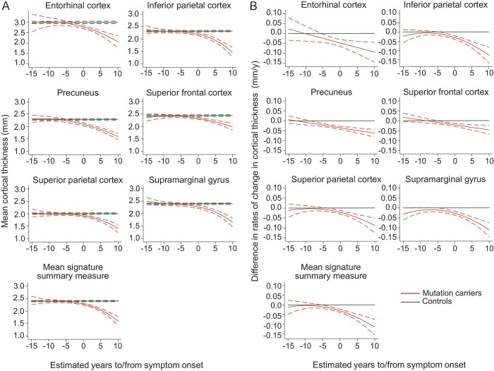
The cortical signature included 6 regions: entorhinal cortex, inferior parietal cortex, precuneus, superior parietal cortex, superior frontal cortex, and supramarginal gyrus. There were significant differences in mean cortical signature thickness between mutation carriers and controls 3 years before predicted symptom onset. The earliest significant difference in a single region, detectable 4 years preonset, was in the precuneus. Rate of change in cortical thickness became significantly different in the cortical signature at 5 years before predicted onset, and in the precuneus at 8 years preonset. Baseline mean signature thickness predicted rate of subsequent thinning and correlated with presymptomatic cognitive change.
The FAD cortical signature appears to be similar to that described for sporadic AD. All component regions showed significant presymptomatic thinning. A composite signature may provide more robust results than a single region and have utility as an outcome measure in presymptomatic trials.
识别家族性阿尔茨海默病(FAD)中皮质变薄的皮质特征模式,并评估其在检测和追踪症状前神经退行性变中的效用。
我们招募了43名FAD突变携带者——36名PSEN1突变携带者、7名APP突变携带者(20名有症状,23名无症状)——以及42名健康对照者,进行纵向临床和MRI研究。所有参与者在基线时均进行了T1加权MRI扫描;55名个体(33名突变携带者;22名对照者)大约每年进行多次(平均2.9次)随访扫描。使用FreeSurfer测量皮质厚度。从有症状的FAD参与者中识别出皮质变薄特征。然后,我们检查了无症状携带者中该特征区域的皮质厚度变化,并评估了其与认知表现的相关性。
皮质特征包括6个区域:内嗅皮质、顶下小叶、楔前叶、顶上小叶、额上回和缘上回。在预测症状出现前3年,突变携带者和对照者之间的平均皮质特征厚度存在显著差异。单个区域最早的显著差异可在发病前4年检测到,位于楔前叶。预测发病前5年,皮质特征区域的皮质厚度变化率显著不同,发病前8年,楔前叶的皮质厚度变化率显著不同。基线平均特征厚度可预测随后的变薄率,并与症状前的认知变化相关。
FAD皮质特征似乎与散发性AD中描述的特征相似。所有组成区域在症状前均出现显著变薄。与单个区域相比,复合特征可能提供更可靠的结果,并可作为症状前试验的一项结局指标。