• 文献检索
  • 文档翻译
  • 深度研究
  • 学术资讯
  • Suppr Zotero 插件Zotero 插件
  • 邀请有礼
  • 套餐&价格
  • 历史记录
应用&插件
Suppr Zotero 插件Zotero 插件浏览器插件Mac 客户端Windows 客户端微信小程序
定价
高级版会员购买积分包购买API积分包
服务
文献检索文档翻译深度研究API 文档MCP 服务
关于我们
关于 Suppr公司介绍联系我们用户协议隐私条款
关注我们

Suppr 超能文献

核心技术专利:CN118964589B侵权必究
粤ICP备2023148730 号-1Suppr @ 2026

文献检索

告别复杂PubMed语法,用中文像聊天一样搜索,搜遍4000万医学文献。AI智能推荐,让科研检索更轻松。

立即免费搜索

文件翻译

保留排版,准确专业,支持PDF/Word/PPT等文件格式,支持 12+语言互译。

免费翻译文档

深度研究

AI帮你快速写综述,25分钟生成高质量综述,智能提取关键信息,辅助科研写作。

立即免费体验

致心律失常性右室心肌病的心室结构:超越容积作为风险衡量指标

Ventricular structure in ARVC: going beyond volumes as a measure of risk.

作者信息

McLeod Kristin, Wall Samuel, Leren Ida Skrinde, Saberniak Jørg, Haugaa Kristina Hermann

机构信息

Cardiac Modelling Department, Simula Research Laboratory, PO Box 134, Oslo, Norway.

Center for Cardiological Innovation, Oslo, Norway.

出版信息

J Cardiovasc Magn Reson. 2016 Oct 14;18(1):73. doi: 10.1186/s12968-016-0291-9.

DOI:10.1186/s12968-016-0291-9
PMID:27756409
原文链接:https://pmc.ncbi.nlm.nih.gov/articles/PMC5069945/
Abstract

BACKGROUND

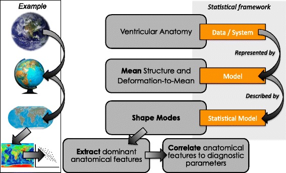
Altered right ventricular structure is an important feature of Arrhythmogenic Right Ventricular Cardiomyopathy (ARVC), but is challenging to quantify objectively. The aim of this study was to go beyond ventricular volumes and diameters and to explore if the shape of the right and left ventricles could be assessed and related to clinical measures. We used quantifiable computational methods to automatically identify and analyse malformations in ARVC patients from Cardiovascular Magnetic Resonance (CMR) images. Furthermore, we investigated how automatically extracted structural features were related to arrhythmic events.

METHODS

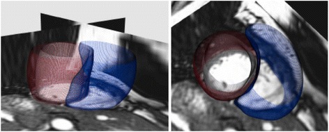
A retrospective cross-sectional feasibility study was performed on CMR short axis cine images of 27 ARVC patients and 21 ageing asymptomatic control subjects. All images were segmented at the end-diastolic (ED) and end-systolic (ES) phases of the cardiac cycle to create three-dimensional (3D) bi-ventricle shape models for each subject. The most common components to single- and bi-ventricular shape in the ARVC population were identified and compared to those obtained from the control group. The correlations were calculated between identified ARVC shapes and parameters from the 2010 Task Force Criteria, in addition to clinical outcomes such as ventricular arrhythmias.

RESULTS

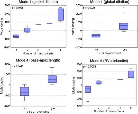
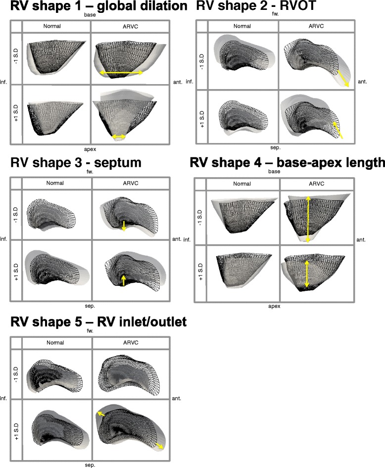
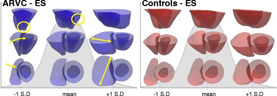
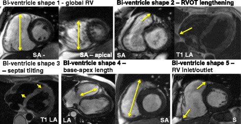
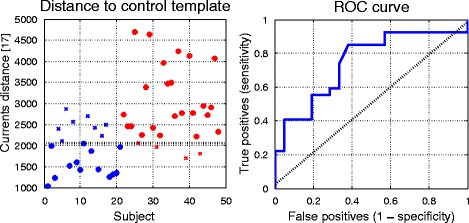
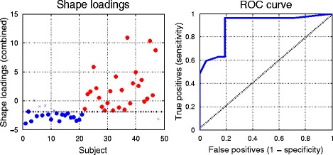
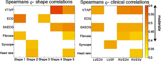
Bi-ventricle shape for the ARVC population showed, as ordered by prevalence with the percent of total variance in the population explained by each shape: global dilation/shrinking of both ventricles (44 %), elongation/shortening at the right ventricle (RV) outflow tract (15 %), tilting at the septum (10 %), shortening/lengthening of both ventricles (7 %), and bulging/shortening at both the RV inflow and outflow (5 %). Bi-ventricle shapes were significantly correlated to several clinical diagnostic parameters and outcomes, including (but not limited to) correlations between global dilation and electrocardiography (ECG) major criteria (p = 0.002), and base-to-apex lengthening and history of arrhythmias (p = 0.003). Classification of ARVC vs. control using shape modes yielded high sensitivity (96 %) and moderate specificity (81 %).

CONCLUSION

We presented for the first time an automatic method for quantifying and analysing ventricular shapes in ARVC patients from CMR images. Specific ventricular shape features were highly correlated with diagnostic indices in ARVC patients and yielded high classification sensitivity. Ventricular shape analysis may be a novel approach to classify ARVC disease, and may be used in diagnosis and in risk stratification for ventricular arrhythmias.

摘要

背景

右心室结构改变是致心律失常性右心室心肌病(ARVC)的一个重要特征,但客观量化具有挑战性。本研究的目的是超越心室容积和直径,探讨是否可以评估右心室和左心室的形状,并将其与临床指标相关联。我们使用可量化的计算方法,从心血管磁共振(CMR)图像中自动识别和分析ARVC患者的畸形情况。此外,我们还研究了自动提取的结构特征与心律失常事件之间的关系。

方法

对27例ARVC患者和21例老年无症状对照者的CMR短轴电影图像进行了一项回顾性横断面可行性研究。在心动周期的舒张末期(ED)和收缩末期(ES)阶段对所有图像进行分割,为每个受试者创建三维(3D)双心室形状模型。确定了ARVC人群中单心室和双心室形状最常见的组成部分,并与对照组获得的组成部分进行比较。除了室性心律失常等临床结果外,还计算了确定的ARVC形状与2010年工作组标准中的参数之间的相关性。

结果

ARVC人群的双心室形状按患病率排序,每种形状在人群中总方差的百分比解释如下:两个心室的整体扩张/收缩(44%)、右心室(RV)流出道的伸长/缩短(15%)、室间隔的倾斜(10%)、两个心室的缩短/伸长(7%)以及RV流入和流出处的膨出/缩短(5%)。双心室形状与几个临床诊断参数和结果显著相关,包括(但不限于)整体扩张与心电图(ECG)主要标准之间的相关性(p = 0.002),以及心底到心尖的延长与心律失常病史之间的相关性(p = 0.003)。使用形状模式对ARVC与对照组进行分类,灵敏度高(96%),特异性中等(81%)。

结论

我们首次提出了一种从CMR图像中量化和分析ARVC患者心室形状的自动方法。特定心室形状特征与ARVC患者的诊断指标高度相关,并具有高分类灵敏度。心室形状分析可能是一种对ARVC疾病进行分类的新方法,可用于诊断和室性心律失常的风险分层。

https://cdn.ncbi.nlm.nih.gov/pmc/blobs/24c2/5069945/724f68e06a23/12968_2016_291_Fig9_HTML.jpg
https://cdn.ncbi.nlm.nih.gov/pmc/blobs/24c2/5069945/a57dac8ff6e6/12968_2016_291_Fig1_HTML.jpg
https://cdn.ncbi.nlm.nih.gov/pmc/blobs/24c2/5069945/51e1c677349d/12968_2016_291_Fig2_HTML.jpg
https://cdn.ncbi.nlm.nih.gov/pmc/blobs/24c2/5069945/ec5437b2229b/12968_2016_291_Fig3_HTML.jpg
https://cdn.ncbi.nlm.nih.gov/pmc/blobs/24c2/5069945/7775dfdbb270/12968_2016_291_Fig4_HTML.jpg
https://cdn.ncbi.nlm.nih.gov/pmc/blobs/24c2/5069945/1dd513c74281/12968_2016_291_Fig5_HTML.jpg
https://cdn.ncbi.nlm.nih.gov/pmc/blobs/24c2/5069945/1eeda6df98dd/12968_2016_291_Fig6_HTML.jpg
https://cdn.ncbi.nlm.nih.gov/pmc/blobs/24c2/5069945/1f765156ccba/12968_2016_291_Fig7_HTML.jpg
https://cdn.ncbi.nlm.nih.gov/pmc/blobs/24c2/5069945/2e11050b8c67/12968_2016_291_Fig8_HTML.jpg
https://cdn.ncbi.nlm.nih.gov/pmc/blobs/24c2/5069945/724f68e06a23/12968_2016_291_Fig9_HTML.jpg

相似文献

1
Ventricular structure in ARVC: going beyond volumes as a measure of risk.致心律失常性右室心肌病的心室结构:超越容积作为风险衡量指标
J Cardiovasc Magn Reson. 2016 Oct 14;18(1):73. doi: 10.1186/s12968-016-0291-9.
2
Long term CMR follow up of patients with right ventricular abnormality and clinically suspected arrhythmogenic right ventricular cardiomyopathy (ARVC).右心室异常且临床疑似致心律失常性右心室心肌病(ARVC)患者的长期 CMR 随访。
J Cardiovasc Magn Reson. 2019 Dec 12;21(1):76. doi: 10.1186/s12968-019-0581-0.
3
Cardiac magnetic resonance based deformation imaging: role of feature tracking in athletes with suspected arrhythmogenic right ventricular cardiomyopathy.基于心脏磁共振的形变成像:特征追踪在疑似致心律失常性右室心肌病运动员中的作用
Int J Cardiovasc Imaging. 2019 Mar;35(3):529-538. doi: 10.1007/s10554-018-1478-y. Epub 2018 Oct 31.
4
Right Ventricular Strain and Dyssynchrony Assessment in Arrhythmogenic Right Ventricular Cardiomyopathy: Cardiac Magnetic Resonance Feature-Tracking Study.致心律失常性右室心肌病的右心室应变与不同步评估:心脏磁共振特征追踪研究
Circ Cardiovasc Imaging. 2015 Nov;8(11):e003647; discussion e003647. doi: 10.1161/CIRCIMAGING.115.003647.
5
Right Ventricular Strain Derived from Cardiac MRI Feature Tracking for the Diagnosis and Prognosis of Arrhythmogenic Right Ventricular Cardiomyopathy.基于心脏 MRI 特征追踪的右心室应变在心律失常性右室心肌病诊断和预后中的价值。
Radiol Cardiothorac Imaging. 2024 Jun;6(3):e230292. doi: 10.1148/ryct.230292.
6
Biventricular myocardial strain analysis using cardiac magnetic resonance feature tracking (CMR-FT) in patients with distinct types of right ventricular diseases comparing arrhythmogenic right ventricular cardiomyopathy (ARVC), right ventricular outflow-tract tachycardia (RVOT-VT), and Brugada syndrome (BrS).使用心脏磁共振特征追踪(CMR-FT)分析不同类型右心室疾病患者的双心室心肌应变,比较致心律失常性右心室心肌病(ARVC)、右心室流出道心动过速(RVOT-VT)和 Brugada 综合征(BrS)。
Clin Res Cardiol. 2019 Oct;108(10):1147-1162. doi: 10.1007/s00392-019-01450-w. Epub 2019 Mar 13.
7
Comparison of patients with early-phase arrhythmogenic right ventricular cardiomyopathy and right ventricular outflow tract ventricular tachycardia.早期致心律失常性右室心肌病患者与右室流出道室性心动过速患者的比较。
Eur Heart J Cardiovasc Imaging. 2017 Jan;18(1):62-69. doi: 10.1093/ehjci/jew014. Epub 2016 Feb 21.
8
Combination of ECG and Echocardiography for Identification of Arrhythmic Events in Early ARVC.心电图和超声心动图联合用于早期 ARVC 心律失常事件的识别。
JACC Cardiovasc Imaging. 2017 May;10(5):503-513. doi: 10.1016/j.jcmg.2016.06.011. Epub 2016 Oct 19.
9
Right ventricular outflow tract dimensions in arrhythmogenic right ventricular cardiomyopathy/dysplasia-a multicentre study comparing echocardiography and cardiovascular magnetic resonance.心律失常性右室心肌病/发育不良的右室流出道的维度:超声心动图与心血管磁共振的多中心研究比较。
Eur Heart J Cardiovasc Imaging. 2018 May 1;19(5):516-523. doi: 10.1093/ehjci/jex092.
10
Right ventricular systolic function and mechanical dispersion identify patients with arrhythmogenic right ventricular cardiomyopathy.右心室收缩功能和机械离散度可识别致心律失常性右心室心肌病患者。
Clin Physiol Funct Imaging. 2018 Sep;38(5):779-787. doi: 10.1111/cpf.12479. Epub 2017 Nov 6.

引用本文的文献

1
Right ventricular function and determining factors of dysfunction in ST-segment-elevation myocardial infarction: a cross-sectional study with cardiac magnetic resonance imaging (MRI).ST段抬高型心肌梗死患者右心室功能及功能障碍的决定因素:一项心脏磁共振成像(MRI)横断面研究
Quant Imaging Med Surg. 2024 Sep 1;14(9):6895-6907. doi: 10.21037/qims-23-1804. Epub 2024 Jun 11.
2
Prognostic value of right atrial strains in arrhythmogenic right ventricular cardiomyopathy.右心房应变在致心律失常性右室心肌病中的预后价值
Insights Imaging. 2024 Feb 27;15(1):58. doi: 10.1186/s13244-024-01630-x.
3
Analysing functional implications of differences in left ventricular morphology using statistical shape modelling.

本文引用的文献

1
A statistical shape modelling framework to extract 3D shape biomarkers from medical imaging data: assessing arch morphology of repaired coarctation of the aorta.一种从医学影像数据中提取三维形状生物标志物的统计形状建模框架:评估修复后的主动脉缩窄的弓部形态
BMC Med Imaging. 2016 May 31;16(1):40. doi: 10.1186/s12880-016-0142-z.
2
Comparison of patients with early-phase arrhythmogenic right ventricular cardiomyopathy and right ventricular outflow tract ventricular tachycardia.早期致心律失常性右室心肌病患者与右室流出道室性心动过速患者的比较。
Eur Heart J Cardiovasc Imaging. 2017 Jan;18(1):62-69. doi: 10.1093/ehjci/jew014. Epub 2016 Feb 21.
3
运用统计形状建模分析左心室形态差异的功能意义。
Sci Rep. 2022 Nov 10;12(1):19163. doi: 10.1038/s41598-022-15888-y.
4
Atlas-based methods for efficient characterization of patient-specific ventricular activation patterns.基于图谱的方法用于高效描述患者特定的心室激活模式。
Europace. 2021 Mar 4;23(23 Suppl 1):i88-i95. doi: 10.1093/europace/euaa397.
5
Cardiac magnetic resonance feature tracking of the right ventricle in convalescent Kawasaki disease in a large single center.恢复期川崎病患者右心室磁共振特征追踪:一项大型单中心研究。
Clin Cardiol. 2021 Jan;44(1):108-115. doi: 10.1002/clc.23512. Epub 2020 Nov 12.
6
Right ventricular shape and function: cardiovascular magnetic resonance reference morphology and biventricular risk factor morphometrics in UK Biobank.右心室形态和功能:英国生物库中心血管磁共振参考形态和双心室危险因素形态计量学
J Cardiovasc Magn Reson. 2019 Jul 18;21(1):41. doi: 10.1186/s12968-019-0551-6.
7
Journal of Cardiovascular Magnetic Resonance 2017.《心血管磁共振杂志》 2017 年刊
J Cardiovasc Magn Reson. 2018 Dec 28;20(1):89. doi: 10.1186/s12968-018-0518-z.
8
Cardiac magnetic resonance based deformation imaging: role of feature tracking in athletes with suspected arrhythmogenic right ventricular cardiomyopathy.基于心脏磁共振的形变成像:特征追踪在疑似致心律失常性右室心肌病运动员中的作用
Int J Cardiovasc Imaging. 2019 Mar;35(3):529-538. doi: 10.1007/s10554-018-1478-y. Epub 2018 Oct 31.
9
Review of Journal of Cardiovascular Magnetic Resonance (JCMR) 2015-2016 and transition of the JCMR office to Boston.回顾《心血管磁共振杂志》(JCMR)2015-2016 年的出版情况以及该杂志办公室迁至波士顿的情况。
J Cardiovasc Magn Reson. 2017 Dec 28;19(1):108. doi: 10.1186/s12968-017-0423-x.
10
Investigating Cardiac Motion Patterns Using Synthetic High-Resolution 3D Cardiovascular Magnetic Resonance Images and Statistical Shape Analysis.使用合成高分辨率3D心血管磁共振图像和统计形状分析研究心脏运动模式。
Front Pediatr. 2017 Mar 8;5:34. doi: 10.3389/fped.2017.00034. eCollection 2017.
Treatment of arrhythmogenic right ventricular cardiomyopathy/dysplasia: an international task force consensus statement.
致心律失常性右室心肌病/发育异常的治疗:一项国际特别工作组共识声明
Eur Heart J. 2015 Dec 7;36(46):3227-37. doi: 10.1093/eurheartj/ehv162. Epub 2015 Jul 27.
4
Recommendations for cardiac chamber quantification by echocardiography in adults: an update from the American Society of Echocardiography and the European Association of Cardiovascular Imaging.超声心动图成人左心室容量和射血分数测量:美国超声心动图学会和欧洲心血管影像协会的更新建议。
J Am Soc Echocardiogr. 2015 Jan;28(1):1-39.e14. doi: 10.1016/j.echo.2014.10.003.
5
Vigorous physical activity impairs myocardial function in patients with arrhythmogenic right ventricular cardiomyopathy and in mutation positive family members.剧烈体育活动会损害致心律失常性右室心肌病患者及突变阳性家庭成员的心肌功能。
Eur J Heart Fail. 2014 Dec;16(12):1337-44. doi: 10.1002/ejhf.181. Epub 2014 Oct 16.
6
Left ventricular shape variation in asymptomatic populations: the Multi-Ethnic Study of Atherosclerosis.无症状人群的左心室形态变化:动脉粥样硬化多族裔研究
J Cardiovasc Magn Reson. 2014 Jul 30;16(1):56. doi: 10.1186/s12968-014-0056-2.
7
Mutation-positive arrhythmogenic right ventricular dysplasia/cardiomyopathy: the triangle of dysplasia displaced.致心律失常性右室发育不良/心肌病:发育不良的三角移位。
J Cardiovasc Electrophysiol. 2013 Dec;24(12):1311-20. doi: 10.1111/jce.12222. Epub 2013 Jul 25.
8
A statistical model for quantification and prediction of cardiac remodelling: application to tetralogy of Fallot.用于心脏重构定量和预测的统计模型:在法洛四联症中的应用。
IEEE Trans Med Imaging. 2011 Sep;30(9):1605-16. doi: 10.1109/TMI.2011.2135375.
9
Right ventricular mechanical dispersion is related to malignant arrhythmias: a study of patients with arrhythmogenic right ventricular cardiomyopathy and subclinical right ventricular dysfunction.右室机械离散度与恶性心律失常相关:心律失常性右室心肌病和亚临床右室功能障碍患者的研究。
Eur Heart J. 2011 May;32(9):1089-96. doi: 10.1093/eurheartj/ehr069. Epub 2011 Mar 15.
10
A review of segmentation methods in short axis cardiac MR images.短轴心脏磁共振图像分割方法综述。
Med Image Anal. 2011 Apr;15(2):169-84. doi: 10.1016/j.media.2010.12.004. Epub 2010 Dec 24.