Biffi Benedetta, Bruse Jan L, Zuluaga Maria A, Ntsinjana Hopewell N, Taylor Andrew M, Schievano Silvia
Centre for Cardiovascular Imaging, UCL Institute of Cardiovascular Science & Great Ormond Street Hospital for Children, London, UK; Department of Medical Physics and Biomedical Engineering, University College London, London, UK.
Centre for Cardiovascular Imaging, UCL Institute of Cardiovascular Science & Great Ormond Street Hospital for Children , London , UK.
Front Pediatr. 2017 Mar 8;5:34. doi: 10.3389/fped.2017.00034. eCollection 2017.
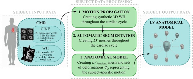
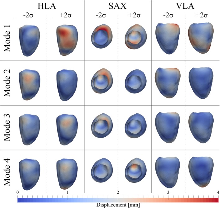
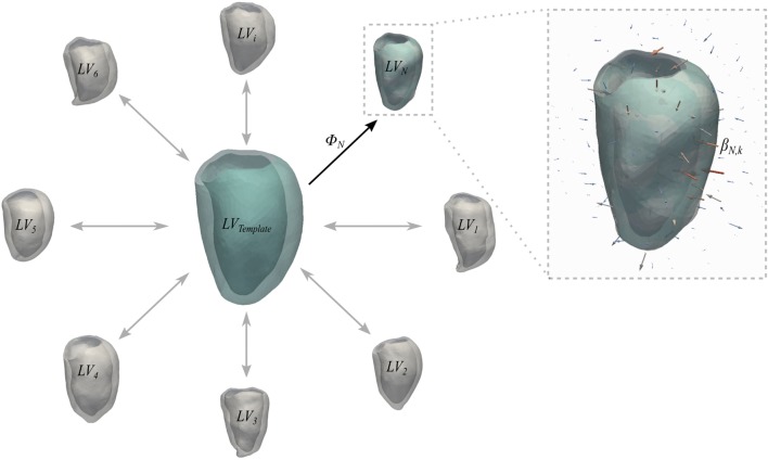
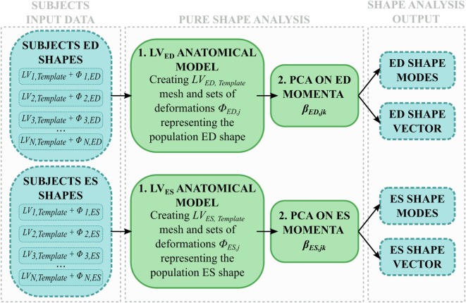
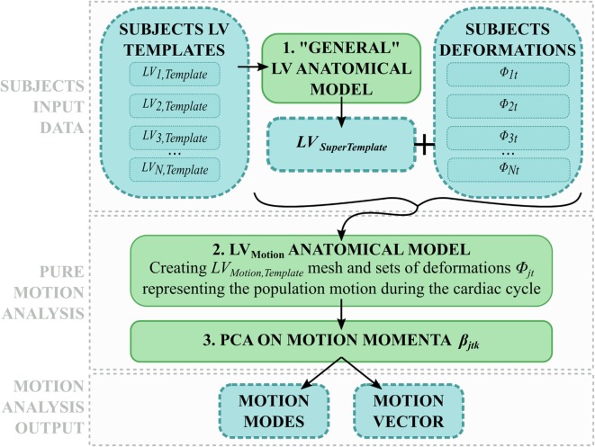
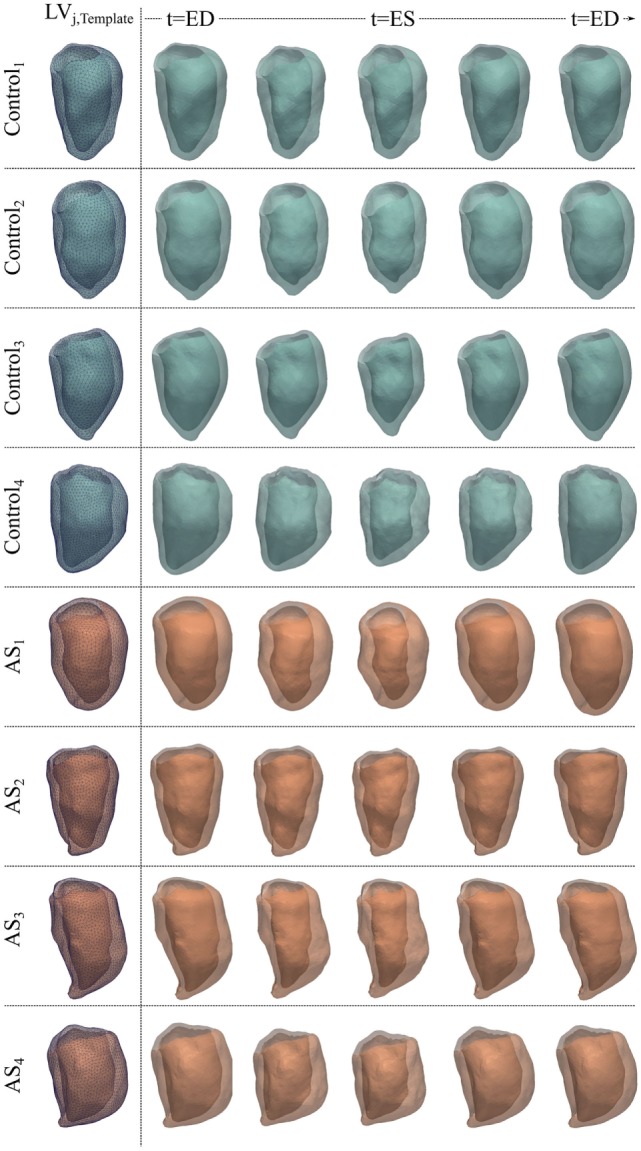
Diagnosis of ventricular dysfunction in congenital heart disease is more and more based on medical imaging, which allows investigation of abnormal cardiac morphology and correlated abnormal function. Although analysis of 2D images represents the clinical standard, novel tools performing automatic processing of 3D images are becoming available, providing more detailed and comprehensive information than simple 2D morphometry. Among these, statistical shape analysis (SSA) allows a consistent and quantitative description of a population of complex shapes, as a way to detect novel biomarkers, ultimately improving diagnosis and pathology understanding. The aim of this study is to describe the implementation of a SSA method for the investigation of 3D left ventricular shape and motion patterns and to test it on a small sample of 4 congenital repaired aortic stenosis patients and 4 age-matched healthy volunteers to demonstrate its potential. The advantage of this method is the capability of analyzing subject-specific motion patterns separately from the individual morphology, visually and quantitatively, as a way to identify functional abnormalities related to both dynamics and shape. Specifically, we combined 3D, high-resolution whole heart data with 2D, temporal information provided by cine cardiovascular magnetic resonance images, and we used an SSA approach to analyze 3D motion . Preliminary results of this pilot study showed that using this method, some differences in end-diastolic and end-systolic ventricular shapes could be captured, but it was not possible to clearly separate the two cohorts based on shape information alone. However, further analyses on ventricular motion allowed to qualitatively identify differences between the two populations. Moreover, by describing shape and motion with a small number of principal components, this method offers a fully automated process to obtain visually intuitive and numerical information on cardiac shape and motion, which could be, once validated on a larger sample size, easily integrated into the clinical workflow. To conclude, in this preliminary work, we have implemented state-of-the-art automatic segmentation and SSA methods, and we have shown how they could improve our understanding of ventricular kinetics by visually and potentially quantitatively highlighting aspects that are usually not picked up by traditional approaches.
先天性心脏病心室功能障碍的诊断越来越依赖医学成像技术,该技术能够对异常的心脏形态及相关的异常功能进行检查。尽管二维图像分析是临床标准,但能够自动处理三维图像的新型工具正逐渐问世,与简单的二维形态测量相比,能提供更详细、全面的信息。其中,统计形状分析(SSA)能够对复杂形状群体进行一致且定量的描述,以此来检测新的生物标志物,最终改善诊断和对病理的理解。本研究的目的是描述一种用于研究三维左心室形状和运动模式的SSA方法的实施过程,并在4例先天性修复主动脉瓣狭窄患者和4例年龄匹配的健康志愿者的小样本上进行测试,以证明其潜力。该方法的优势在于能够将个体特定的运动模式与个体形态分开进行视觉和定量分析,从而识别与动力学和形状相关的功能异常。具体而言,我们将三维高分辨率全心数据与二维电影心血管磁共振图像提供的时间信息相结合,并使用SSA方法分析三维运动。这项初步研究的结果表明,使用该方法可以捕捉到舒张末期和收缩末期心室形状的一些差异,但仅基于形状信息无法清晰区分这两组人群。然而,对心室运动的进一步分析能够定性地识别出这两个人群之间的差异。此外,通过用少量主成分描述形状和运动,该方法提供了一个完全自动化的过程,以获取关于心脏形状和运动的直观视觉和数值信息,一旦在更大样本量上得到验证,就可以很容易地整合到临床工作流程中。总之,在这项初步工作中,我们实施了最先进的自动分割和SSA方法,并展示了它们如何通过视觉上和潜在的定量方式突出传统方法通常无法捕捉到的方面,从而提高我们对心室动力学的理解。