Lewis Joshua B, Milner Dallin C, Lewis Adam L, Dunaway Todd M, Egbert Kaleb M, Albright Scott C, Merrell Brigham J, Monson Troy D, Broberg Dallin S, Gassman Jason R, Thomas Daniel B, Arroyo Juan A, Reynolds Paul R
Lung and Placenta Research Laboratory, Physiology and Developmental Biology, Brigham Young University, Provo, UT 84602, USA.
Int J Environ Res Public Health. 2016 Oct 17;13(10):1018. doi: 10.3390/ijerph13101018.
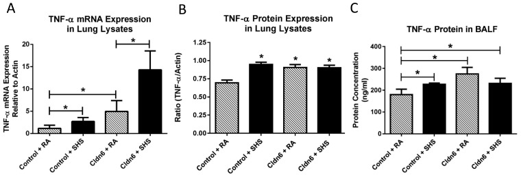
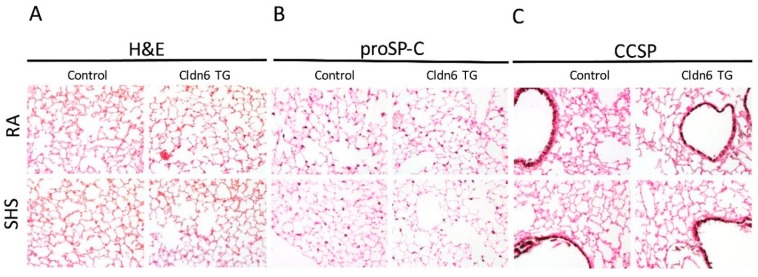
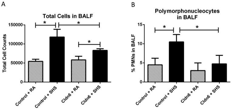
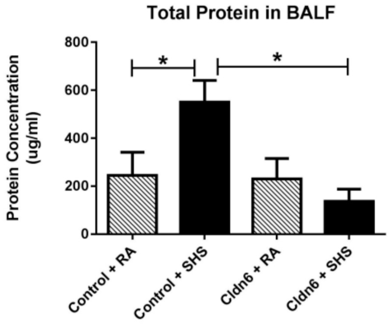
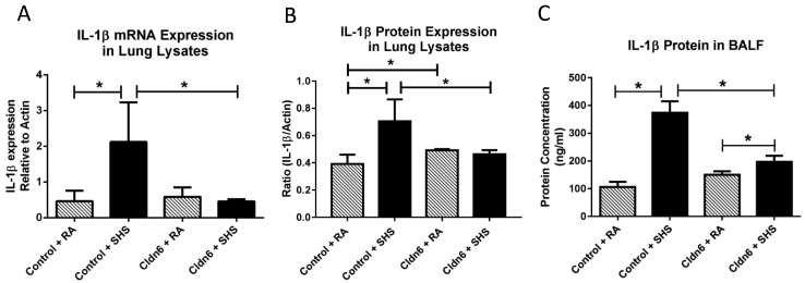
It has long been understood that increased epithelial permeability contributes to inflammation observed in many respiratory diseases. Recently, evidence has revealed that environmental exposure to noxious material such as cigarette smoke reduces tight junction barrier integrity, thus enhancing inflammatory conditions. Claudin-6 (Cldn6) is a tetraspanin transmembrane protein found within the tight junctional complex and is implicated in maintaining lung epithelial barriers. To test the hypothesis that increased Cldn6 ameliorates inflammation at the respiratory barrier, we utilized the Tet-On inducible transgenic system to conditionally over-express Clnd6 in the distal lung. Cldn6 transgenic (TG) and control mice were continuously provided doxycycline from postnatal day (PN) 30 until euthanasia date at PN90. A subset of Cldn6 TG and control mice were also subjected to daily secondhand tobacco smoke (SHS) via a nose only inhalation system from PN30-90 and compared to room air (RA) controls. Animals were euthanized on PN90 and lungs were harvested for histological and molecular characterization. Bronchoalveolar lavage fluid (BALF) was procured for the assessment of inflammatory cells and molecules. Quantitative RT-PCR and immunoblotting revealed increased Cldn6 expression in TG vs. control animals and SHS decreased Cldn6 expression regardless of genetic up-regulation. Histological evaluations revealed no adverse pulmonary remodeling via Hematoxylin and Eosin (H&E) staining or any qualitative alterations in the abundance of type II pneumocytes or proximal non-ciliated epithelial cells via staining for cell specific propeptide of Surfactant Protein-C (proSP-C) or Club Cell Secretory Protein (CCSP), respectively. Immunoblotting and qRT-PCR confirmed the differential expression of Cldn6 and the pro-inflammatory cytokines TNF-α and IL-1β. As a general theme, inflammation induced by SHS exposure was influenced by the availability of Cldn6. These data reveal captivating information suggesting a role for Cldn6 in lungs exposed to tobacco smoke. Further research is critically necessary in order to fully explain roles for tight junctional components such as Cldn6 and other related molecules in lungs coping with exposure.
长期以来,人们一直认为上皮通透性增加会导致许多呼吸道疾病中出现的炎症。最近,有证据表明,暴露于香烟烟雾等有害物质的环境会降低紧密连接屏障的完整性,从而加剧炎症状态。Claudin-6(Cldn6)是一种四跨膜蛋白,存在于紧密连接复合物中,与维持肺上皮屏障有关。为了验证增加Cldn6可改善呼吸道屏障炎症这一假设,我们利用Tet-On诱导转基因系统在肺远端条件性过表达Clnd6。从出生后第30天(PN30)到PN90安乐死之日,持续给Cldn6转基因(TG)小鼠和对照小鼠提供强力霉素。从PN30到PN90,还通过仅鼻吸入系统让一部分Cldn6 TG小鼠和对照小鼠每天接触二手烟(SHS),并与室内空气(RA)对照进行比较。在PN90对动物实施安乐死,并采集肺组织进行组织学和分子特征分析。获取支气管肺泡灌洗液(BALF)以评估炎症细胞和分子。定量逆转录聚合酶链反应(qRT-PCR)和免疫印迹显示,与对照动物相比,TG动物中Cldn6表达增加,且无论基因上调情况如何,SHS都会降低Cldn6表达。组织学评估显示,苏木精和伊红(H&E)染色未发现不良肺重塑,通过分别对表面活性蛋白C(proSP-C)或俱乐部细胞分泌蛋白(CCSP)的细胞特异性前体肽进行染色,II型肺细胞或近端无纤毛上皮细胞的丰度也无任何定性改变。免疫印迹和qRT-PCR证实了Cldn6以及促炎细胞因子肿瘤坏死因子-α(TNF-α)和白细胞介素-1β(IL-1β)的差异表达。总体而言,SHS暴露诱导的炎症受Cldn6可用性的影响。这些数据揭示了引人入胜的信息,表明Cldn6在暴露于烟草烟雾的肺中发挥作用。为了充分解释紧密连接成分如Cldn6和其他相关分子在应对暴露的肺中的作用,进一步的研究至关重要。