Long Niamh M, Plodkowski Andrew J, Schor-Bardach Rachel, Geyer Alexander I, Zheng Junting, Moskowitz Chaya S, Ginsberg Michelle S
From the Departments of *Radiology, †Medicine, and ‡Epidemiology and Biostatistics, Memorial Sloan Kettering Cancer Center, New York, NY.
J Comput Assist Tomogr. 2017 May/Jun;41(3):437-441. doi: 10.1097/RCT.0000000000000520.
The aims of this study were to describe the computed tomographic features of organizing pneumonia (OP) in an oncologic patient population and to also identify features associated with lung cancer and patients undergoing hematopoietic stem cell transplant (HSCT).
In retrospective computed tomographies from 151 patients with pathologically confirmed OP between January 2009 and September 2014, number of lesions, location, size, margin type, and consistency, as well as volume of lymphadenopathy and the presence and size of pleural effusions, were recorded. Associated malignancy was noted.
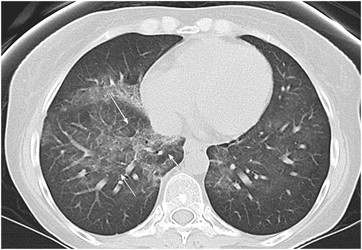
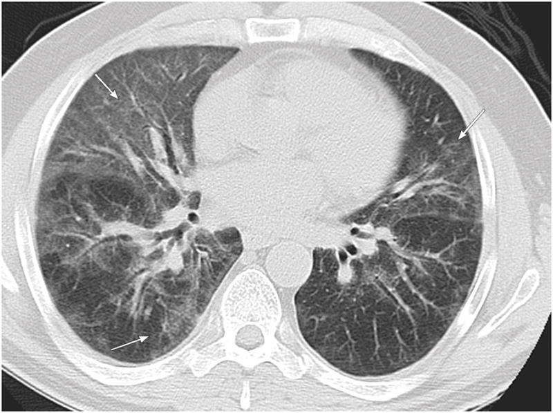
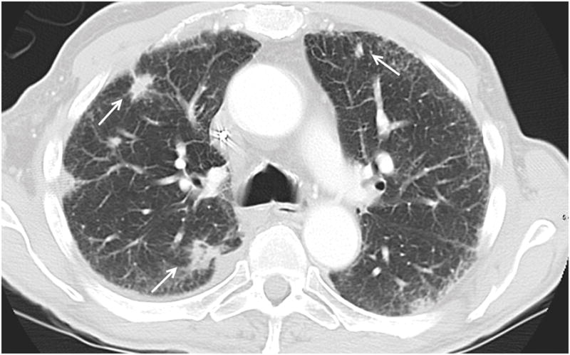
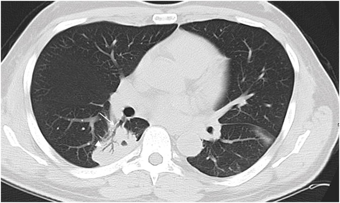
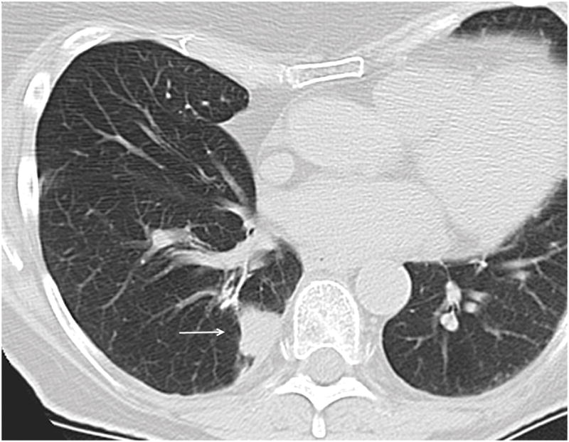
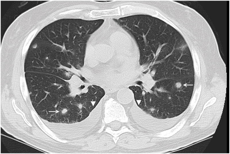
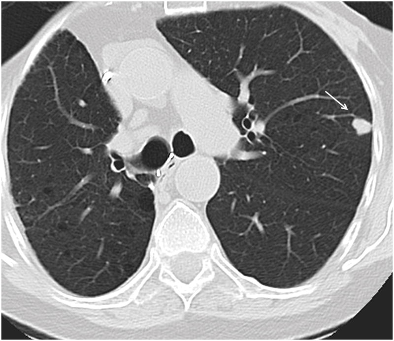
Organizing pneumonia most commonly presented as a diffuse process (n = 62, 41%), frequently occupied both a central and peripheral location (n = 79, 53%), and commonly presented with a solid appearance (n = 67, 44%) or with ground glass opacity (n = 80, 53%). Pleural effusions were seen in 68 patients (45%). Organizing pneumonia less frequently contained air bronchograms, cavitation, necrosis, surrounding ground glass opacity, or adjacent bronchiectasis. In patients with lung cancer (n = 25, 17%), OP more likely presented as discrete lesions and occupied a peripheral location as compared with patients with other malignancies (Ps = 0.025 and 0.002). In HSCT patients (n = 29, 19%), a diffuse process was more commonly seen than in non-HSCT patients (P = 0.038).
Organizing pneumonia more commonly presents as discrete lesions with a peripheral location in patients with lung cancer and as a diffuse process in patients who had undergone HSCT.
本研究旨在描述肿瘤患者群体中机化性肺炎(OP)的计算机断层扫描特征,并确定与肺癌以及接受造血干细胞移植(HSCT)患者相关的特征。
回顾性分析2009年1月至2014年9月期间151例经病理证实为OP患者的计算机断层扫描结果,记录病变数量、位置、大小、边缘类型、密度,以及淋巴结肿大的体积、胸腔积液的有无及大小。记录相关的恶性肿瘤情况。
机化性肺炎最常表现为弥漫性病变(n = 62,41%),常同时累及中央和周边部位(n = 79,53%),常见表现为实性(n = 67,44%)或磨玻璃影(n = 80,53%)。68例患者(45%)可见胸腔积液。机化性肺炎较少出现空气支气管征、空洞、坏死、周围磨玻璃影或邻近支气管扩张。与其他恶性肿瘤患者相比,肺癌患者(n = 25,17%)的OP更可能表现为孤立性病变且位于周边部位(P值分别为0.025和0.002)。在HSCT患者(n = 29,19%)中,弥漫性病变比非HSCT患者更常见(P = 0.038)。
机化性肺炎在肺癌患者中更常表现为周边部位的孤立性病变,在接受HSCT的患者中更常表现为弥漫性病变。