R. Dimbleby Department of Cancer Research, Randall Division of Cell and Molecular Biophysics, Division of Cancer Studies, King's College London , Guy's Campus New Hunt's House, London SE1 1UL, U.K.
Department of Imaging Chemistry and Biology, Division of Imaging Sciences and Biomedical Engineering, St. Thomas' Hospital, King's College London , London SE1 7EH, U.K.
ACS Nano. 2017 Jan 24;11(1):249-257. doi: 10.1021/acsnano.6b05356. Epub 2016 Nov 3.
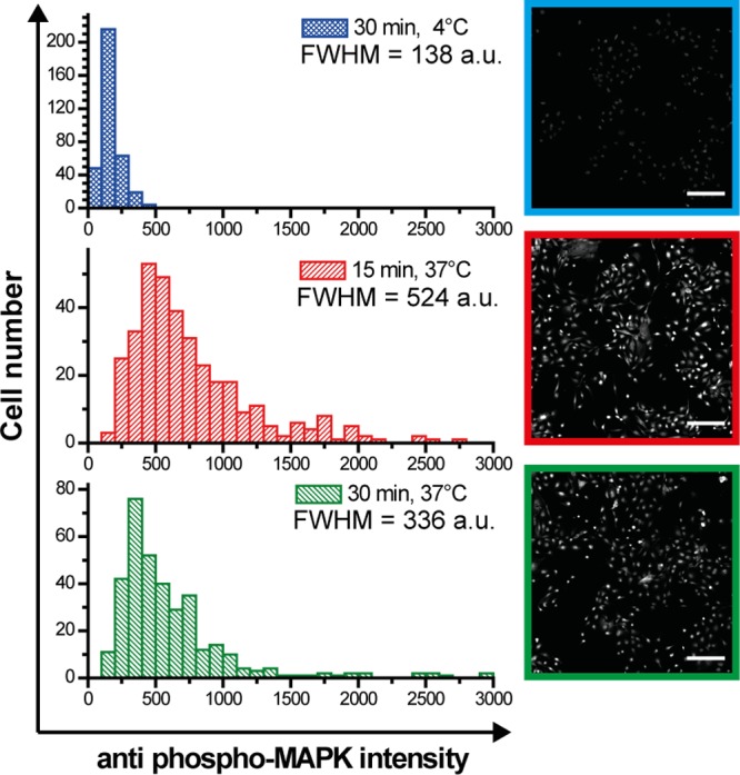
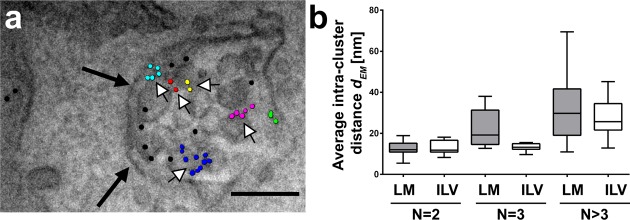
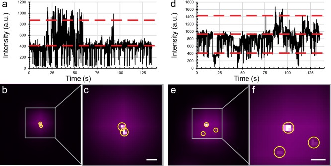
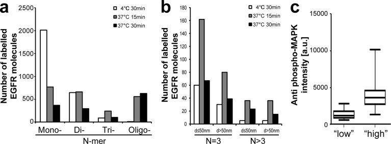
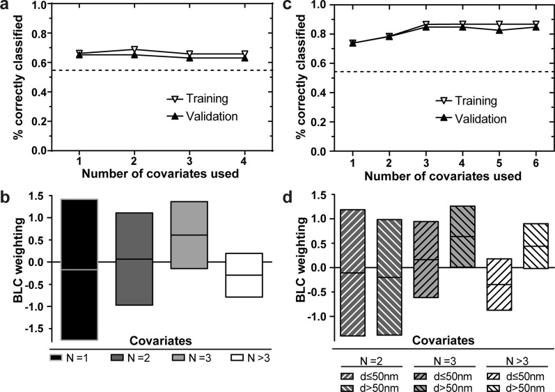
Heterogeneity of mitogen-activated protein kinase (MAPK) activation in genetically identical cells, which occurs in response to epidermal growth factor receptor (EGFR) signaling, remains poorly understood. MAPK cascades integrate signals emanating from different EGFR spatial locations, including the plasma membrane and endocytic compartment. We previously hypothesized that in EGF-stimulated cells the MAPK phosphorylation (pMAPK) level and activity are largely determined by the spatial organization of the EGFR clusters within the cell. For experimental testing of this hypothesis, we used super-resolution microscopy to define EGFR clusters by receptor numbers (N) and average intracluster distances (d). From these data, we predicted the extent of pMAPK with 85% accuracy on a cell-to-cell basis with control data returning 54% accuracy (P < 0.001). For comparison, the prediction accuracy was only 61% (P = 0.382) when the diffraction-limited averaged fluorescence intensity/cluster was used. Large clusters (N ≥ 3) with d > 50 nm were most predictive for pMAPK level in cells. Electron microscopy revealed that these large clusters were primarily localized to the limiting membrane of multivesicular bodies (MVB). Many tighter packed dimers/multimers (d < 50 nm) were found on intraluminal vesicles within MVBs, where they were unlikely to activate MAPK because of the physical separation. Our results suggest that cell-to-cell differences in N and d contain crucial information to predict EGFR-activated cellular pMAPK levels and explain pMAPK heterogeneity in isogenic cells.
不同细胞内的丝裂原活化蛋白激酶(MAPK)在受到表皮生长因子受体(EGFR)信号刺激时会发生激活,但其异质性目前还知之甚少。MAPK 级联反应整合了来自不同 EGFR 空间位置的信号,包括质膜和内吞隔室。我们之前假设,在 EGF 刺激的细胞中,MAPK 磷酸化(pMAPK)的水平和活性在很大程度上取决于细胞内 EGFR 簇的空间组织。为了实验验证这一假设,我们使用超分辨率显微镜根据受体数量(N)和平均簇内距离(d)来定义 EGFR 簇。根据这些数据,我们可以在细胞间基础上以 85%的准确度预测 pMAPK,而对照数据的准确度仅为 54%(P < 0.001)。相比之下,当使用衍射受限的平均荧光强度/簇时,预测准确性仅为 61%(P = 0.382)。具有 d > 50nm 的大簇(N≥3)对于预测细胞内 pMAPK 水平最具预测性。电子显微镜显示,这些大簇主要定位于多泡体(MVB)的限界膜。在 MVB 内的腔内小泡中发现了许多更紧密的二聚体/多聚体(d < 50nm),由于物理分离,它们不太可能激活 MAPK。我们的研究结果表明,细胞间 N 和 d 的差异包含了预测 EGFR 激活的细胞内 pMAPK 水平的关键信息,并解释了同基因细胞中 pMAPK 的异质性。