Lindqvist Richard, Mundt Filip, Gilthorpe Jonathan D, Wölfel Silke, Gekara Nelson O, Kröger Andrea, Överby Anna K
Department of Clinical Microbiology, Virology, Umeå University, 90185, Umeå, Sweden.
The Laboratory for Molecular Infection Medicine Sweden (MIMS), 90187, Umeå, Sweden.
J Neuroinflammation. 2016 Oct 24;13(1):277. doi: 10.1186/s12974-016-0748-7.
Neurotropic flaviviruses such as tick-borne encephalitis virus (TBEV), Japanese encephalitis virus (JEV), West Nile virus (WNV), and Zika virus (ZIKV) are causative agents of severe brain-related diseases including meningitis, encephalitis, and microcephaly. We have previously shown that local type I interferon response within the central nervous system (CNS) is involved in the protection of mice against tick-borne flavivirus infection. However, the cells responsible for mounting this protective response are not defined.
Primary astrocytes were isolated from wild-type (WT) and interferon alpha receptor knock out (IFNAR) mice and infected with neurotropic flaviviruses. Viral replication and spread, IFN induction and response, and cellular viability were analyzed. Transcriptional levels in primary astrocytes treated with interferon or supernatant from virus-infected cells were analyzed by RNA sequencing and evaluated by different bioinformatics tools.
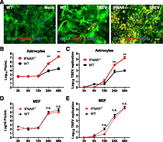
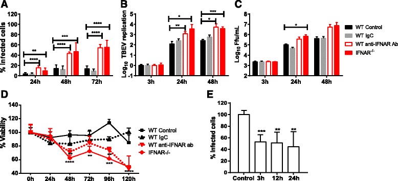
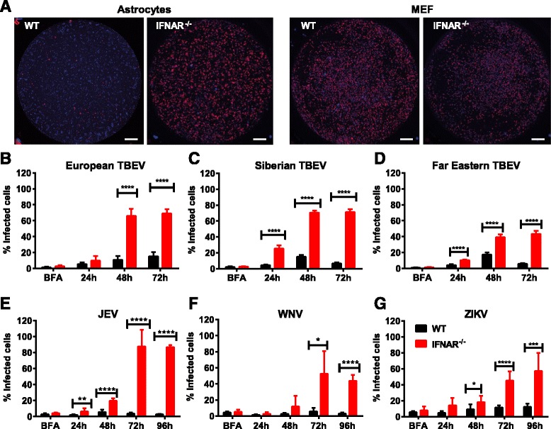
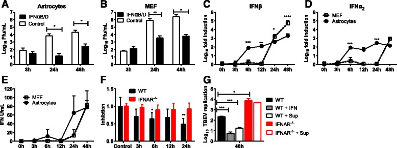
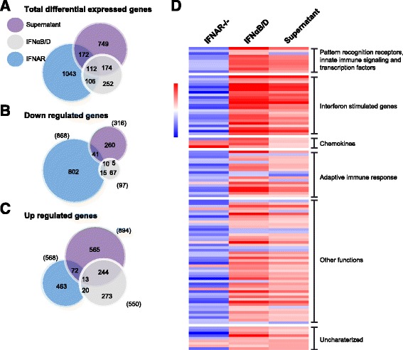
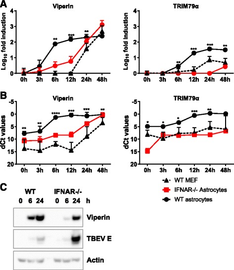
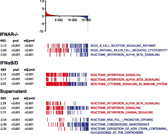
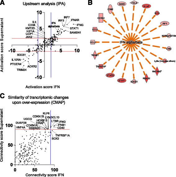
Here, we show that astrocytes control viral replication of different TBEV strains, JEV, WNV, and ZIKV. In contrast to fibroblast, astrocytes mount a rapid interferon response and restrict viral spread. Furthermore, basal expression levels of key interferon-stimulated genes are high in astrocytes compared to mouse embryonic fibroblasts. Bioinformatic analysis of RNA-sequencing data reveals that astrocytes have established a basal antiviral state which contributes to the rapid viral recognition and upregulation of interferons. The most highly upregulated pathways in neighboring cells were linked to type I interferon response and innate immunity. The restriction in viral growth was dependent on interferon signaling, since loss of the interferon receptor, or its blockade in wild-type cells, resulted in high viral replication and virus-induced cytopathic effects. Astrocyte supernatant from TBEV-infected cells can restrict TBEV growth in astrocytes already 6 h post infection, the effect on neurons is highly reinforced, and astrocyte supernatant from 3 h post infection is already protective.
These findings suggest that the combination of an intrinsic constitutive antiviral response and the fast induction of type I IFN production by astrocytes play an important role in self-protection of astrocytes and suppression of flavivirus replication in the CNS.
嗜神经性黄病毒,如蜱传脑炎病毒(TBEV)、日本脑炎病毒(JEV)、西尼罗河病毒(WNV)和寨卡病毒(ZIKV),是包括脑膜炎、脑炎和小头畸形在内的严重脑部相关疾病的病原体。我们之前已经表明,中枢神经系统(CNS)内的局部I型干扰素反应参与了小鼠抵抗蜱传黄病毒感染的保护过程。然而,引发这种保护反应的细胞尚未明确。
从野生型(WT)和干扰素α受体敲除(IFNAR)小鼠中分离出原代星形胶质细胞,并感染嗜神经性黄病毒。分析病毒复制与传播、干扰素诱导与反应以及细胞活力。通过RNA测序分析用干扰素或病毒感染细胞的上清液处理的原代星形胶质细胞中的转录水平,并通过不同的生物信息学工具进行评估。
在此,我们表明星形胶质细胞可控制不同TBEV毒株、JEV、WNV和ZIKV的病毒复制。与成纤维细胞不同,星形胶质细胞能迅速产生干扰素反应并限制病毒传播。此外,与小鼠胚胎成纤维细胞相比,星形胶质细胞中关键干扰素刺激基因的基础表达水平较高。对RNA测序数据的生物信息学分析表明,星形胶质细胞已建立了基础抗病毒状态,这有助于快速识别病毒并上调干扰素。相邻细胞中上调程度最高的通路与I型干扰素反应和固有免疫相关。病毒生长的限制依赖于干扰素信号传导,因为干扰素受体缺失或在野生型细胞中对其进行阻断会导致病毒大量复制和病毒诱导的细胞病变效应。TBEV感染细胞的星形胶质细胞上清液在感染后6小时就能限制TBEV在星形胶质细胞中的生长,对神经元的作用得到高度增强,感染后3小时的星形胶质细胞上清液就已具有保护作用。
这些发现表明,星形胶质细胞固有的组成性抗病毒反应与快速诱导产生I型干扰素的结合,在星形胶质细胞的自我保护和抑制CNS中黄病毒复制方面发挥着重要作用。