• 文献检索
  • 文档翻译
  • 深度研究
  • 学术资讯
  • Suppr Zotero 插件Zotero 插件
  • 邀请有礼
  • 套餐&价格
  • 历史记录
应用&插件
Suppr Zotero 插件Zotero 插件浏览器插件Mac 客户端Windows 客户端微信小程序
定价
高级版会员购买积分包购买API积分包
服务
文献检索文档翻译深度研究API 文档MCP 服务
关于我们
关于 Suppr公司介绍联系我们用户协议隐私条款
关注我们

Suppr 超能文献

核心技术专利:CN118964589B侵权必究
粤ICP备2023148730 号-1Suppr @ 2026

文献检索

告别复杂PubMed语法,用中文像聊天一样搜索,搜遍4000万医学文献。AI智能推荐,让科研检索更轻松。

立即免费搜索

文件翻译

保留排版,准确专业,支持PDF/Word/PPT等文件格式,支持 12+语言互译。

免费翻译文档

深度研究

AI帮你快速写综述,25分钟生成高质量综述,智能提取关键信息,辅助科研写作。

立即免费体验

使用免费软件Echo E-waves对舒张功能进行运动学分析——可行性与可重复性

Kinematic analysis of diastolic function using the freely available software Echo E-waves - feasibility and reproducibility.

作者信息

Sundqvist Martin G, Salman Katrin, Tornvall Per, Ugander Martin

机构信息

Department of Clinical Science and Education, Södersjukhuset, Karolinska Institutet, and Cardiology Clinic, Södersjukhuset, SE-188 83, Stockholm, Sweden.

Department of Clinical Physiology, Karolinska Institutet, and Karolinska University Hospital, SE-171 76, Stockholm, Sweden.

出版信息

BMC Med Imaging. 2016 Oct 27;16(1):60. doi: 10.1186/s12880-016-0162-8.

DOI:10.1186/s12880-016-0162-8
PMID:27784288
原文链接:https://pmc.ncbi.nlm.nih.gov/articles/PMC5082357/
Abstract

BACKGROUND

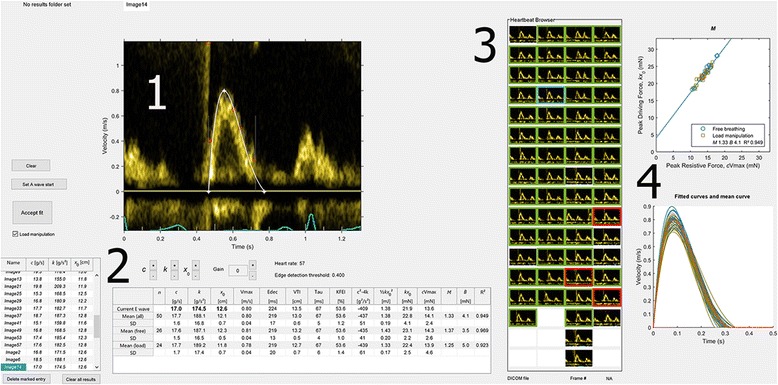
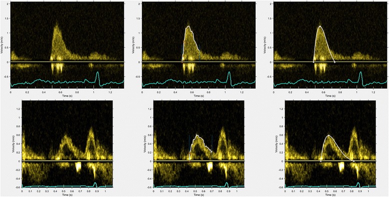
Early diastolic left ventricular (LV) filling can be accurately described using the same methods used in classical mechanics to describe the motion of a loaded spring as it recoils, a validated method also referred to as the Parameterized Diastolic Filling (PDF) formalism. With this method, each E-wave recorded by pulsed wave (PW) Doppler can be mathematically described in terms of three constants: LV stiffness (k), viscoelasticity (c), and load (x ). Also, additional parameters of physiological and diagnostic interest can be derived. An efficient software application for PDF analysis has not been available. We aim to describe the structure, feasibility, time efficiency and intra-and interobserver variability for use of such a solution, implemented in Echo E-waves, a freely available software application ( www.echoewaves.org ).

RESULTS

An application was developed, with the ability to open DICOM files from different vendors, as well as rapid semi-automatic analysis and export of results. E-waves from 20 patients were analyzed by two investigators. Analysis time for a median of 34 (interquartile range (IQR) 29-42) E-waves per patient (representing 63 %, IQR 56-79 % of the recorded E-waves per patient) was 4.3 min (IQR 4.0-4.6 min). Intra-and intraobserver variability was good or excellent for 12 out of 14 parameters (coefficient of variation 2.5-18.7 %, intraclass correlation coefficient 0.80-0.99).

CONCLUSION

Kinematic analysis of diastolic function using the PDF method for Doppler echocardiography implemented in freely available semiautomatic software is highly feasible, time efficient, and has good to excellent intra-and interobserver variability.

摘要

背景

舒张早期左心室(LV)充盈可以使用经典力学中描述加载弹簧回弹运动的相同方法进行准确描述,这种经过验证的方法也称为参数化舒张期充盈(PDF)形式。使用这种方法,脉冲波(PW)多普勒记录的每个E波都可以用三个常数进行数学描述:左心室僵硬度(k)、粘弹性(c)和负荷(x)。此外,还可以得出具有生理和诊断意义的其他参数。目前尚未有用于PDF分析的高效软件应用程序。我们旨在描述一种在免费软件应用程序Echo E-waves(www.echoewaves.org)中实现的此类解决方案的结构、可行性、时间效率以及观察者内和观察者间的变异性。

结果

开发了一个应用程序,它能够打开来自不同供应商的DICOM文件,并能进行快速半自动分析和结果导出。两名研究人员对20名患者的E波进行了分析。每位患者中位数为34个(四分位间距(IQR)为29 - 42个)E波的分析时间(占每位患者记录的E波的63%,IQR为56 - 79%)为4.3分钟(IQR为4.0 - 4.6分钟)。14个参数中的12个参数的观察者内和观察者间变异性良好或极佳(变异系数为2.5 - 18.7%,组内相关系数为0.80 - 0.99)。

结论

在免费的半自动软件中使用PDF方法对多普勒超声心动图的舒张功能进行运动学分析是高度可行、时间高效的,并且观察者内和观察者间变异性良好或极佳。

https://cdn.ncbi.nlm.nih.gov/pmc/blobs/3fb1/5082357/5266d46e390b/12880_2016_162_Fig2_HTML.jpg
https://cdn.ncbi.nlm.nih.gov/pmc/blobs/3fb1/5082357/077bb54fb4d7/12880_2016_162_Fig1_HTML.jpg
https://cdn.ncbi.nlm.nih.gov/pmc/blobs/3fb1/5082357/5266d46e390b/12880_2016_162_Fig2_HTML.jpg
https://cdn.ncbi.nlm.nih.gov/pmc/blobs/3fb1/5082357/077bb54fb4d7/12880_2016_162_Fig1_HTML.jpg
https://cdn.ncbi.nlm.nih.gov/pmc/blobs/3fb1/5082357/5266d46e390b/12880_2016_162_Fig2_HTML.jpg

相似文献

1
Kinematic analysis of diastolic function using the freely available software Echo E-waves - feasibility and reproducibility.使用免费软件Echo E-waves对舒张功能进行运动学分析——可行性与可重复性
BMC Med Imaging. 2016 Oct 27;16(1):60. doi: 10.1186/s12880-016-0162-8.
2
Normal Reference Values for Assessing Diastolic Function Using the Parameterized Diastolic Filling Formalism Method in Patients with Normal Results of Rest and Stress Echocardiography.在静息和负荷超声心动图结果正常的患者中,使用参数化舒张期充盈形式法评估舒张功能的正常参考值。
Ultrasound Med Biol. 2018 Nov;44(11):2261-2266. doi: 10.1016/j.ultrasmedbio.2018.06.014. Epub 2018 Jul 31.
3
Quantifying Diastolic Function: From E-Waves as Triangles to Physiologic Contours via the 'Geometric Method'.舒张功能的量化:从将E波视为三角形到通过“几何方法”得出生理轮廓。
Cardiovasc Eng Technol. 2018 Mar;9(1):105-119. doi: 10.1007/s13239-017-0339-5. Epub 2018 Jan 16.
4
The kinematic filling efficiency index of the left ventricle: contrasting normal vs. diabetic physiology.左心室的运动学充盈效率指数:正常生理与糖尿病生理的对比
Ultrasound Med Biol. 2007 Jun;33(6):842-50. doi: 10.1016/j.ultrasmedbio.2006.11.003. Epub 2007 May 3.
5
Vortex formation time-to-left ventricular early rapid filling relation: model-based prediction with echocardiographic validation.涡旋形成时间与左心室早期快速充盈的关系:基于模型的预测与超声心动图验证。
J Appl Physiol (1985). 2010 Dec;109(6):1812-9. doi: 10.1152/japplphysiol.00645.2010. Epub 2010 Sep 23.
6
Fractionating E-wave deceleration time into its stiffness and relaxation components distinguishes pseudonormal from normal filling.将 E 波减速时间进行僵硬度和弛豫成分的分段,可将假性正常充盈与正常充盈区分开来。
Circ Cardiovasc Imaging. 2015 Jan;8(1). doi: 10.1161/CIRCIMAGING.114.002177.
7
Interstudy reproducibility of parameters of left ventricular diastolic function: a Doppler echocardiography study.左心室舒张功能参数的研究间可重复性:一项多普勒超声心动图研究。
J Am Soc Echocardiogr. 2003 Nov;16(11):1128-35. doi: 10.1067/S0894-7317(03)00641-2.
8
Reproducibility of Doppler echocardiographic assessment of left ventricular diastolic function in multicenter setting.多中心环境下左心室舒张功能的多普勒超声心动图评估的可重复性。
J Am Soc Echocardiogr. 2005 Feb;18(2):99-106. doi: 10.1016/j.echo.2004.08.003.
9
Assessment of left ventricular diastolic function by gated single-photon emission tomography: comparison with Doppler echocardiography.门控单光子发射断层扫描评估左心室舒张功能:与多普勒超声心动图的比较。
Eur J Nucl Med Mol Imaging. 2003 Nov;30(11):1532-7. doi: 10.1007/s00259-003-1303-1. Epub 2003 Aug 21.
10
Reproducibility and inter-vendor variability of left ventricular deformation measurements by three-dimensional speckle-tracking echocardiography.三维斑点追踪超声心动图测量左心室变形的可重复性和不同仪器间的可变性。
J Am Soc Echocardiogr. 2011 Aug;24(8):878-85. doi: 10.1016/j.echo.2011.04.016. Epub 2011 Jun 8.

引用本文的文献

1
The size of myocardial infarction and peri-infarction edema are not major determinants of diastolic impairment after acute myocardial infarction.心肌梗死的面积和梗死周围水肿并非急性心肌梗死后舒张功能障碍的主要决定因素。
Int J Cardiovasc Imaging. 2025 Jan;41(1):103-112. doi: 10.1007/s10554-024-03294-6. Epub 2024 Dec 5.
2
Diastolic function and its association with diabetes, hypertension and age in an outpatient population with normal stress echocardiography findings.在静息超声心动图检查正常的门诊人群中,舒张功能及其与糖尿病、高血压和年龄的关系。
Cardiovasc Ultrasound. 2020 Nov 20;18(1):46. doi: 10.1186/s12947-020-00228-9.
3

本文引用的文献

1
Recommendations for the Evaluation of Left Ventricular Diastolic Function by Echocardiography: An Update from the American Society of Echocardiography and the European Association of Cardiovascular Imaging.超声心动图评估左心室舒张功能的建议:美国超声心动图学会和欧洲心血管影像学会的更新
J Am Soc Echocardiogr. 2016 Apr;29(4):277-314. doi: 10.1016/j.echo.2016.01.011.
2
Quantification of global diastolic function by kinematic modeling-based analysis of transmitral flow via the parametrized diastolic filling formalism.通过基于运动学建模的经二尖瓣血流分析,采用参数化舒张期充盈形式对整体舒张功能进行量化。
J Vis Exp. 2014 Sep 1(91):e51471. doi: 10.3791/51471.
3
Mechanistic validation of the 2016 American Society of Echocardiography/European Association of Cardiovascular Imaging Guidelines for the assessment of diastolic dysfunction in heart failure with reduced ejection fraction.
2016 年美国超声心动图学会/欧洲心血管影像协会心力衰竭伴射血分数降低的舒张功能障碍评估指南的机制学验证。
Cardiovasc Ultrasound. 2020 Oct 16;18(1):42. doi: 10.1186/s12947-020-00224-z.
4
Alternative diastolic function models of ventricular longitudinal filling velocity are mathematically identical.替代的心室纵向充盈速度的舒张功能模型在数学上是相同的。
Am J Physiol Heart Circ Physiol. 2020 May 1;318(5):H1059-H1067. doi: 10.1152/ajpheart.00681.2019. Epub 2020 Mar 6.
5
The link between exercise and titin passive stiffness.运动与肌联蛋白被动僵硬度之间的联系。
Exp Physiol. 2017 Sep 1;102(9):1055-1066. doi: 10.1113/EP086275. Epub 2017 Jul 31.
The kinematic filling efficiency index of the left ventricle: contrasting normal vs. diabetic physiology.
左心室的运动学充盈效率指数:正常生理与糖尿病生理的对比
Ultrasound Med Biol. 2007 Jun;33(6):842-50. doi: 10.1016/j.ultrasmedbio.2006.11.003. Epub 2007 May 3.
4
Load-independent index of diastolic filling: model-based derivation with in vivo validation in control and diastolic dysfunction subjects.舒张期充盈的负荷独立指数:基于模型的推导及在对照和舒张功能障碍受试者中的体内验证
J Appl Physiol (1985). 2006 Jul;101(1):92-101. doi: 10.1152/japplphysiol.01305.2005. Epub 2006 Mar 30.
5
Diabetes and diastolic function: stiffness and relaxation from transmitral flow.糖尿病与舒张功能:经二尖瓣血流的僵硬度与舒张功能
Ultrasound Med Biol. 2005 Dec;31(12):1589-96. doi: 10.1016/j.ultrasmedbio.2005.07.015.
6
The peak atrioventricular pressure gradient to transmitral flow relation: kinematic model prediction with in vivo validation.经二尖瓣血流的峰值房室压力阶差关系:体内验证的运动学模型预测
J Am Soc Echocardiogr. 2004 Aug;17(8):839-44. doi: 10.1016/j.echo.2004.04.017.
7
Recommendations for quantification of Doppler echocardiography: a report from the Doppler Quantification Task Force of the Nomenclature and Standards Committee of the American Society of Echocardiography.多普勒超声心动图定量分析建议:美国超声心动图学会命名与标准委员会多普勒定量工作组报告
J Am Soc Echocardiogr. 2002 Feb;15(2):167-84. doi: 10.1067/mje.2002.120202.
8
Chamber properties from transmitral flow: prediction of average and passive left ventricular diastolic stiffness.经二尖瓣血流的腔室特性:平均和被动左心室舒张期僵硬度的预测
J Appl Physiol (1985). 2001 Jul;91(1):154-62. doi: 10.1152/jappl.2001.91.1.154.
9
Prognostic value of diastolic filling parameters derived using a novel image processing technique in patients > or = 70 years of age with congestive heart failure.
Am J Cardiol. 1999 Jul 1;84(1):82-6. doi: 10.1016/s0002-9149(99)00196-4.
10
Can trasmitral Doppler E-waves differentiate hypertensive hearts from normal?
Hypertension. 1997 Oct;30(4):788-95. doi: 10.1161/01.hyp.30.4.788.