• 文献检索
  • 文档翻译
  • 深度研究
  • 学术资讯
  • Suppr Zotero 插件Zotero 插件
  • 邀请有礼
  • 套餐&价格
  • 历史记录
应用&插件
Suppr Zotero 插件Zotero 插件浏览器插件Mac 客户端Windows 客户端微信小程序
定价
高级版会员购买积分包购买API积分包
服务
文献检索文档翻译深度研究API 文档MCP 服务
关于我们
关于 Suppr公司介绍联系我们用户协议隐私条款
关注我们

Suppr 超能文献

核心技术专利:CN118964589B侵权必究
粤ICP备2023148730 号-1Suppr @ 2026

文献检索

告别复杂PubMed语法,用中文像聊天一样搜索,搜遍4000万医学文献。AI智能推荐,让科研检索更轻松。

立即免费搜索

文件翻译

保留排版,准确专业,支持PDF/Word/PPT等文件格式,支持 12+语言互译。

免费翻译文档

深度研究

AI帮你快速写综述,25分钟生成高质量综述,智能提取关键信息,辅助科研写作。

立即免费体验

脉络膜与光学相干断层扫描

The Choroid and Optical Coherence Tomography.

作者信息

Sezer Taha, Altınışık Muhammet, Koytak İbrahim Arif, Özdemir Mehmet Hakan

机构信息

Bezmialem Vakıf University Faculty of Medicine, Department of Ophthalmology, İstanbul, Turkey.

出版信息

Turk J Ophthalmol. 2016 Jan;46(1):30-37. doi: 10.4274/tjo.10693. Epub 2016 Jan 5.

DOI:10.4274/tjo.10693
PMID:27800255
原文链接:https://pmc.ncbi.nlm.nih.gov/articles/PMC5076307/
Abstract

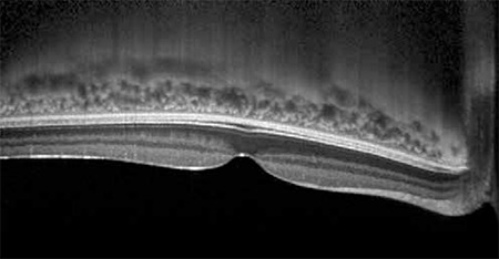
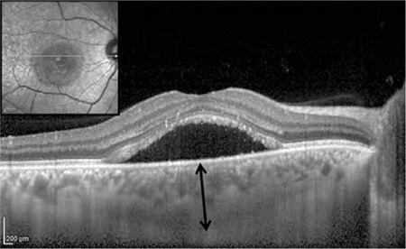
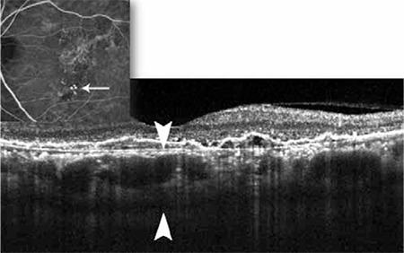
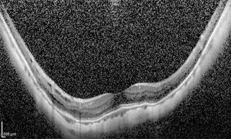
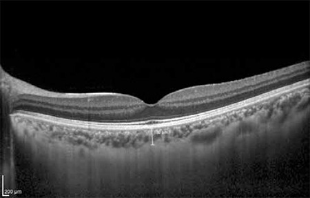
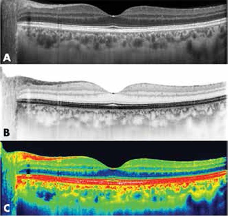
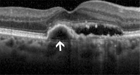
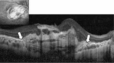
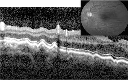
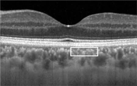
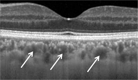
The choroid is the most vascular tissue in the eye and it plays an important role in the pathophysiology of various common chorioretinal diseases such as central serous retinopathy, age-related macular degeneration and degenerative myopia. Quantitative assessment of the choroid has been quite challenging with traditional imaging modalities such as indocyanine green angiography and ultrasonography due to limited resolution and repeatability. With the advent of optical coherence tomography (OCT) technology, detailed visualization of the choroid in vivo is now possible. Measurements of choroidal thickness have also enabled new directions in research to study normal and pathological processes within the choroid. The aim of the present study is to review the current literature on choroidal imaging using OCT.

摘要

脉络膜是眼部血管最丰富的组织,在多种常见的脉络膜视网膜疾病(如中心性浆液性视网膜病变、年龄相关性黄斑变性和变性近视)的病理生理学中发挥着重要作用。由于分辨率和可重复性有限,使用传统成像方式(如吲哚菁绿血管造影和超声检查)对脉络膜进行定量评估颇具挑战性。随着光学相干断层扫描(OCT)技术的出现,现在可以在体内对脉络膜进行详细可视化。脉络膜厚度的测量也为研究脉络膜内正常和病理过程的新研究方向提供了可能。本研究的目的是综述目前关于使用OCT进行脉络膜成像的文献。

https://cdn.ncbi.nlm.nih.gov/pmc/blobs/1254/5076307/c6b3982e4e23/TJO-46-30-g11.jpg
https://cdn.ncbi.nlm.nih.gov/pmc/blobs/1254/5076307/86ee0c73360e/TJO-46-30-g1.jpg
https://cdn.ncbi.nlm.nih.gov/pmc/blobs/1254/5076307/a6d8a5927ceb/TJO-46-30-g2.jpg
https://cdn.ncbi.nlm.nih.gov/pmc/blobs/1254/5076307/fc381b4e6842/TJO-46-30-g3.jpg
https://cdn.ncbi.nlm.nih.gov/pmc/blobs/1254/5076307/93114fc763b3/TJO-46-30-g4.jpg
https://cdn.ncbi.nlm.nih.gov/pmc/blobs/1254/5076307/5e019c748f38/TJO-46-30-g5.jpg
https://cdn.ncbi.nlm.nih.gov/pmc/blobs/1254/5076307/3a21b097c8eb/TJO-46-30-g6.jpg
https://cdn.ncbi.nlm.nih.gov/pmc/blobs/1254/5076307/98770f54bf79/TJO-46-30-g7.jpg
https://cdn.ncbi.nlm.nih.gov/pmc/blobs/1254/5076307/88c2b7d18ab8/TJO-46-30-g8.jpg
https://cdn.ncbi.nlm.nih.gov/pmc/blobs/1254/5076307/f4d37416913e/TJO-46-30-g9.jpg
https://cdn.ncbi.nlm.nih.gov/pmc/blobs/1254/5076307/cf1f56f84292/TJO-46-30-g10.jpg
https://cdn.ncbi.nlm.nih.gov/pmc/blobs/1254/5076307/c6b3982e4e23/TJO-46-30-g11.jpg

相似文献

1
The Choroid and Optical Coherence Tomography.脉络膜与光学相干断层扫描
Turk J Ophthalmol. 2016 Jan;46(1):30-37. doi: 10.4274/tjo.10693. Epub 2016 Jan 5.
2
Investigating the choriocapillaris and choroidal vasculature with new optical coherence tomography technologies.利用新型光学相干断层扫描技术研究脉络膜毛细血管和脉络膜血管。
Prog Retin Eye Res. 2016 May;52:130-55. doi: 10.1016/j.preteyeres.2015.10.002. Epub 2015 Oct 23.
3
Optical coherence tomography: imaging of the choroid and beyond.光学相干断层扫描:脉络膜成像及其他。
Surv Ophthalmol. 2013 Sep-Oct;58(5):387-429. doi: 10.1016/j.survophthal.2012.12.001. Epub 2013 Aug 2.
4
Clinical applications of choroidal imaging technologies.脉络膜成像技术的临床应用
Indian J Ophthalmol. 2015 May;63(5):384-90. doi: 10.4103/0301-4738.159861.
5
Enhanced depth imaging-OCT of the choroid: a review of the current literature.脉络膜增强深度成像光学相干断层扫描:当前文献综述
Graefes Arch Clin Exp Ophthalmol. 2014 Dec;252(12):1871-83. doi: 10.1007/s00417-014-2840-y. Epub 2014 Nov 4.
6
Assessment of macular choroidal thickness by optical coherence tomography and angiographic changes in central serous chorioretinopathy.光学相干断层扫描评估中心性浆液性脉络膜视网膜病变的脉络膜视网膜厚度及造影改变。
Ophthalmology. 2012 Aug;119(8):1666-78. doi: 10.1016/j.ophtha.2012.02.021. Epub 2012 Apr 21.
7
Segregation of ophthalmoscopic characteristics according to choroidal thickness in patients with early age-related macular degeneration.根据早期年龄相关性黄斑变性患者脉络膜厚度对眼底特征进行分离。
Retina. 2012 Jul;32(7):1265-71. doi: 10.1097/IAE.0b013e31824453ac.
8
Imaging chorioretinal vascular disease.眼底血管疾病影像学
Eye (Lond). 2010 Mar;24(3):422-7. doi: 10.1038/eye.2009.309. Epub 2009 Dec 11.
9
OCT angiography and evaluation of the choroid and choroidal vascular disorders.光学相干断层扫描血管造影及脉络膜和脉络膜血管病变的评估。
Prog Retin Eye Res. 2018 Nov;67:30-55. doi: 10.1016/j.preteyeres.2018.07.002. Epub 2018 Jul 27.
10
Morphologic features of focal choroidal excavation on spectral domain optical coherence tomography with simultaneous angiography.频域光学相干断层扫描联合血管造影术观察局限性脉络膜凹陷的形态学特征
Retina. 2014 Jul;34(7):1407-14. doi: 10.1097/IAE.0000000000000108.

引用本文的文献

1
Deep learning-based segmentation of OCT images for choroidal thickness.基于深度学习的光学相干断层扫描(OCT)图像脉络膜厚度分割
J Optom. 2025 Apr-Jun;18(2):100556. doi: 10.1016/j.optom.2025.100556. Epub 2025 May 5.
2
Contrast-enhanced near-infrared photoacoustic microscopy and optical coherence tomography imaging of rat fundus.大鼠眼底的对比增强近红外光声显微镜和光学相干断层扫描成像
Nanophotonics. 2024 Jun 13;13(19):3631-3646. doi: 10.1515/nanoph-2023-0872. eCollection 2024 Aug.
3
How is the posterior ocular segment affected in cesarean section and normal vaginal delivery?

本文引用的文献

1
Choroidal Thickness in Open-angle Glaucoma.开角型青光眼的脉络膜厚度
J Glaucoma. 2015 Oct-Nov;24(8):619-23. doi: 10.1097/IJG.0000000000000063.
2
Peripapillary and macular choroidal thickness in glaucoma.青光眼患者视乳头周围及黄斑区脉络膜厚度
J Ophthalmic Vis Res. 2014 Apr;9(2):154-61.
3
Glaucoma and choroidal thickness.青光眼与脉络膜厚度
剖宫产和正常阴道分娩如何影响后眼部?
Indian J Ophthalmol. 2024 Nov 1;72(Suppl 5):S893-S901. doi: 10.4103/IJO.IJO_1317_24. Epub 2024 Oct 25.
4
The Haemodialysis Session Effect on the Choroidal Thickness and Retinal and Choroidal Microcirculation-A Literature Review.血液透析疗程对脉络膜厚度及视网膜和脉络膜微循环的影响——文献综述
J Clin Med. 2023 Dec 16;12(24):7729. doi: 10.3390/jcm12247729.
5
Evaluation of Choroidal Vascularity Index in Keratoconus Patients: Does Choroidal Vascularity Change in Keratoconus?圆锥角膜患者脉络膜血管指数的评估:圆锥角膜患者的脉络膜血管会发生变化吗?
J Curr Ophthalmol. 2023 Aug 11;35(1):36-41. doi: 10.4103/joco.joco_189_22. eCollection 2023 Jan-Mar.
6
Retinal astrocytic hamartoma complicated by branch retinal vein occlusion in a patient with tuberous sclerosis complex.结节性硬化症患者视网膜星形细胞错构瘤合并视网膜分支静脉阻塞
Am J Ophthalmol Case Rep. 2023 Aug 24;32:101920. doi: 10.1016/j.ajoc.2023.101920. eCollection 2023 Dec.
7
The impact of obesity on ocular hemodynamics and choroidal thickness.肥胖对眼部血流动力学和脉络膜厚度的影响。
Ther Adv Ophthalmol. 2023 Jun 30;15:25158414231180985. doi: 10.1177/25158414231180985. eCollection 2023 Jan-Dec.
8
Three-Dimensional Ultrasound Elastography Detects Age-Related Increase in Anterior Peripapillary Sclera and Optic Nerve Head Compression During IOP Elevation.三维超声弹性成像检测到眼压升高期间前巩膜和视乳头周围神经鞘的年龄相关性增加和视神经头受压。
Invest Ophthalmol Vis Sci. 2023 Jun 1;64(7):16. doi: 10.1167/iovs.64.7.16.
9
Effect of Nasal Septum Deviation on the Choroidal Thickness.鼻中隔偏曲对脉络膜厚度的影响。
Cureus. 2023 Apr 19;15(4):e37840. doi: 10.7759/cureus.37840. eCollection 2023 Apr.
10
Microvascular and structural analysis of the retina and choroid in heart failure patients with reduced ejection fraction.心力衰竭患者射血分数降低的视网膜和脉络膜微血管及结构分析。
Sci Rep. 2023 Apr 4;13(1):5467. doi: 10.1038/s41598-023-32751-w.
J Ophthalmic Vis Res. 2014 Apr;9(2):151-3.
4
Effect of anti-VEGF treatment on choroidal thickness over time in patients with neovascular age-related macular degeneration.抗血管内皮生长因子治疗对新生血管性年龄相关性黄斑变性患者脉络膜厚度随时间的影响。
Eur J Ophthalmol. 2014 Nov-Dec;24(6):897-903. doi: 10.5301/ejo.5000509. Epub 2014 Jul 18.
5
Choroidal thickness in patients with diabetic retinopathy.糖尿病视网膜病变患者的脉络膜厚度
Clin Ophthalmol. 2014 Mar 27;8:637-42. doi: 10.2147/OPTH.S59395. eCollection 2014.
6
Relationship between retinal lesions and inward choroidal bulging in Vogt-Koyanagi-Harada disease.Vogt-小柳-原田病的视网膜病变与脉络膜内陷的关系。
Am J Ophthalmol. 2014 May;157(5):1056-63. doi: 10.1016/j.ajo.2014.01.015. Epub 2014 Jan 31.
7
Analysis of choroidal morphologic features and vasculature in healthy eyes using spectral-domain optical coherence tomography.利用频域光学相干断层扫描分析健康眼的脉络膜形态特征和血管。
Ophthalmology. 2013 Sep;120(9):1901-8. doi: 10.1016/j.ophtha.2013.01.066. Epub 2013 May 9.
8
Optical coherence tomography--current and future applications.光学相干断层扫描——当前和未来的应用。
Curr Opin Ophthalmol. 2013 May;24(3):213-21. doi: 10.1097/ICU.0b013e32835f8bf8.
9
Enhanced depth imaging optical coherence tomography of the choroid in Vogt-Koyanagi-Harada disease.Vogt-小柳原田病脉络膜的增强深度成像光相干断层扫描。
Retina. 2012 Nov-Dec;32(10):2061-9. doi: 10.1097/IAE.0b013e318256205a.
10
Choroidal thickness and visual acuity in highly myopic eyes.高度近视眼中的脉络膜厚度与视力
Retina. 2012 Jul;32(7):1229-36. doi: 10.1097/IAE.0b013e318242b990.