Lavinsky Fabio, Lavinsky Daniel
Department of Ophthalmology, Paulista School of Medicine, São Paulo Hospital, Federal University of São Paulo, São Paulo, Brazil ; Department of Ophthalmology, Federal University of Rio Grande do Sul, Porto Alegre, Brazil ; Department of Ophthalmology, Instituto de Oftalmologia Lavinsky, Rua Quintino Bocaiuva 673, Porto Alegre, RS 90410-140 Brazil.
Department of Ophthalmology, Federal University of Rio Grande do Sul, Porto Alegre, Brazil.
Int J Retina Vitreous. 2016 Nov 1;2:25. doi: 10.1186/s40942-016-0050-y. eCollection 2016.
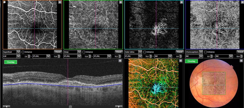
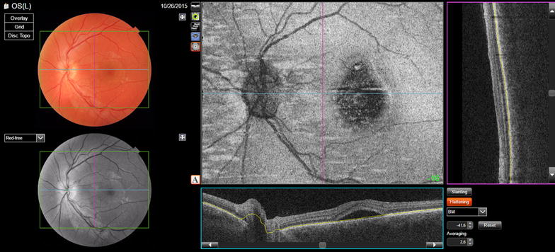
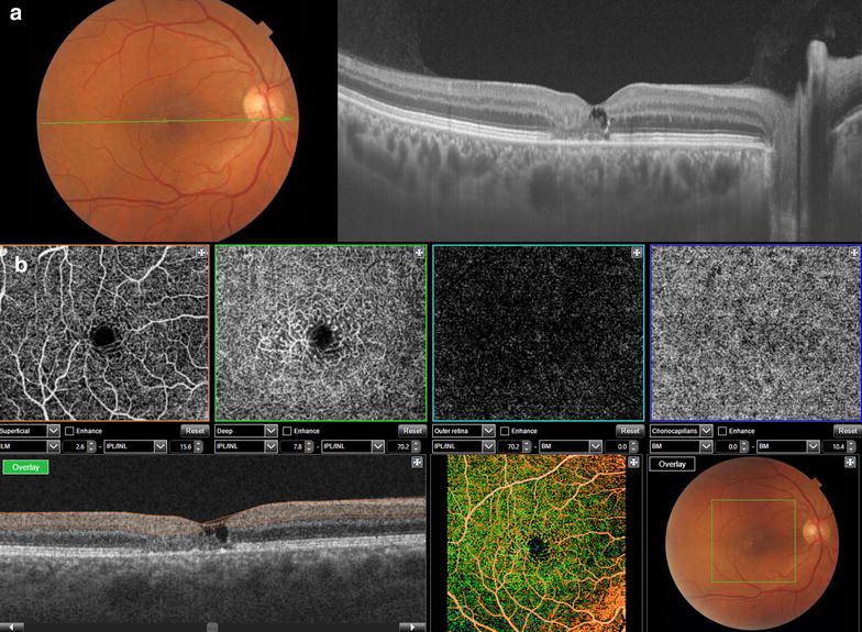
Technologies for multimodal digital imaging of vitreoretinal diseases have improved the accuracy of diagnosis and the depth of the knowledge of the mechanisms of disease and their response to treatments. Optic coherence tomography (OCT) has become a mandatory tool for the management and for the follow-up of retinal pathologies. OCT technology evolved in the last two decades from time-domain to spectral domain and recently to the swept-source OCTs (SS-OCT). SS-OCT improved the depth of imaging and the scan speed, thus adding novel algorithms and features such as for vitreous and vitreoretinal interface evaluation, choroid segmentation and mapping, OCT angiography and En-face OCT. The multimodal approach using SS-OCT is expected to advance the understanding of retinal pathologies such as age related macular degeneration, diabetic maculopathy, central serous chorioretinopathy, the pachychoroid spectrum and macular telangiectasia. Surgical vitreoretinal diseases such as vitreo-macular traction syndrome, epiretinal membrane, retinal detachment, proliferative vitreoretinal retinopathy and diabetic traction retinal detachment also will be better understood and documented with SS-OCT. This technology also provides great utility for a broad spectrum of ophthalmic pathologies including glaucoma, uveitis, tumors and anterior segment evaluation.
玻璃体视网膜疾病的多模态数字成像技术提高了诊断的准确性,加深了对疾病机制及其对治疗反应的认识。光学相干断层扫描(OCT)已成为视网膜疾病管理和随访的必备工具。在过去二十年中,OCT技术从时域发展到频域,最近又发展到扫频源OCT(SS-OCT)。SS-OCT提高了成像深度和扫描速度,从而增加了诸如玻璃体和玻璃体视网膜界面评估、脉络膜分割和测绘、OCT血管造影和表面OCT等新算法和功能。使用SS-OCT的多模态方法有望促进对视网膜疾病的理解,如年龄相关性黄斑变性、糖尿病性黄斑病变、中心性浆液性脉络膜视网膜病变、厚脉络膜谱系和黄斑毛细血管扩张症。诸如玻璃体黄斑牵引综合征、视网膜前膜、视网膜脱离、增殖性玻璃体视网膜病变和糖尿病性牵引性视网膜脱离等手术性玻璃体视网膜疾病,也将通过SS-OCT得到更好的理解和记录。这项技术还为包括青光眼、葡萄膜炎、肿瘤和眼前节评估在内的广泛眼科疾病提供了很大的实用价值。