• 文献检索
  • 文档翻译
  • 深度研究
  • 学术资讯
  • Suppr Zotero 插件Zotero 插件
  • 邀请有礼
  • 套餐&价格
  • 历史记录
应用&插件
Suppr Zotero 插件Zotero 插件浏览器插件Mac 客户端Windows 客户端微信小程序
定价
高级版会员购买积分包购买API积分包
服务
文献检索文档翻译深度研究API 文档MCP 服务
关于我们
关于 Suppr公司介绍联系我们用户协议隐私条款
关注我们

Suppr 超能文献

核心技术专利:CN118964589B侵权必究
粤ICP备2023148730 号-1Suppr @ 2026

文献检索

告别复杂PubMed语法,用中文像聊天一样搜索,搜遍4000万医学文献。AI智能推荐,让科研检索更轻松。

立即免费搜索

文件翻译

保留排版,准确专业,支持PDF/Word/PPT等文件格式,支持 12+语言互译。

免费翻译文档

深度研究

AI帮你快速写综述,25分钟生成高质量综述,智能提取关键信息,辅助科研写作。

立即免费体验

7T 神经影像学的主要临床获益。

Key clinical benefits of neuroimaging at 7T.

机构信息

High Field MR Center, Department of Biomedical Imaging and Image-guided Therapy, Medical University of Vienna, Lazarettgasse 14, A-1090, Vienna, Austria; Christian Doppler Laboratory for Clinical Molecular MRI, Vienna, Austria.

High Field MR Center, Department of Biomedical Imaging and Image-guided Therapy, Medical University of Vienna, Lazarettgasse 14, A-1090, Vienna, Austria.

出版信息

Neuroimage. 2018 Mar;168:477-489. doi: 10.1016/j.neuroimage.2016.11.031. Epub 2016 Nov 13.

DOI:10.1016/j.neuroimage.2016.11.031
PMID:27851995
原文链接:https://pmc.ncbi.nlm.nih.gov/articles/PMC5832016/
Abstract

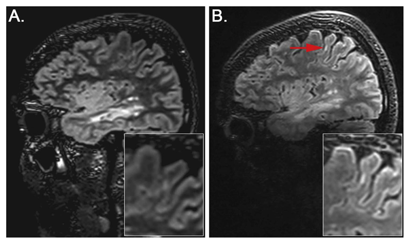
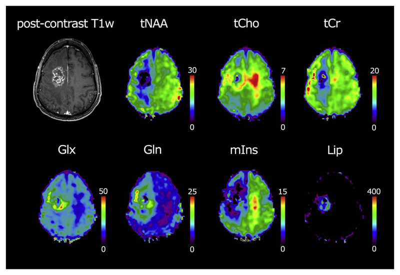
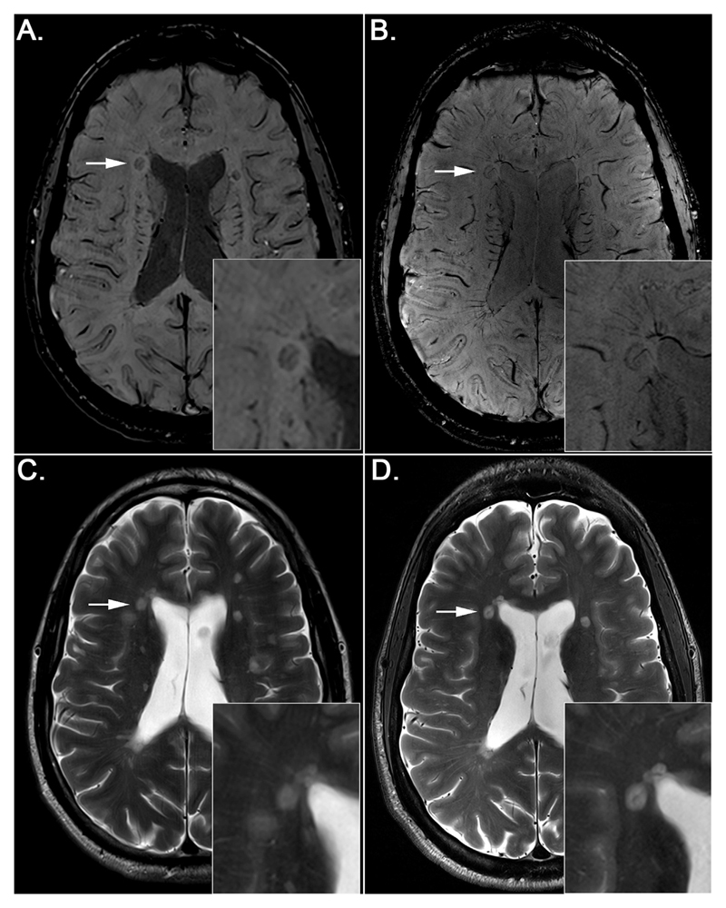
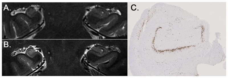
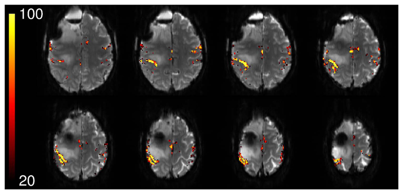
The growing interest in ultra-high field MRI, with more than 35.000 MR examinations already performed at 7T, is related to improved clinical results with regard to morphological as well as functional and metabolic capabilities. Since the signal-to-noise ratio increases with the field strength of the MR scanner, the most evident application at 7T is to gain higher spatial resolution in the brain compared to 3T. Of specific clinical interest for neuro applications is the cerebral cortex at 7T, for the detection of changes in cortical structure, like the visualization of cortical microinfarcts and cortical plaques in Multiple Sclerosis. In imaging of the hippocampus, even subfields of the internal hippocampal anatomy and pathology may be visualized with excellent spatial resolution. Using Susceptibility Weighted Imaging, the plaque-vessel relationship and iron accumulations in Multiple Sclerosis can be visualized, which may provide a prognostic factor of disease. Vascular imaging is a highly promising field for 7T which is dealt with in a separate dedicated article in this special issue. The static and dynamic blood oxygenation level-dependent contrast also increases with the field strength, which significantly improves the accuracy of pre-surgical evaluation of vital brain areas before tumor removal. Improvement in acquisition and hardware technology have also resulted in an increasing number of MR spectroscopic imaging studies in patients at 7T. More recent parallel imaging and short-TR acquisition approaches have overcome the limitations of scan time and spatial resolution, thereby allowing imaging matrix sizes of up to 128×128. The benefits of these acquisition approaches for investigation of brain tumors and Multiple Sclerosis have been shown recently. Together, these possibilities demonstrate the feasibility and advantages of conducting routine diagnostic imaging and clinical research at 7T.

摘要

超高场 MRI 的日益普及,已经有超过 35000 次的 7T 磁共振检查,这与形态学以及功能和代谢能力的临床改善结果有关。由于信号与噪声比随磁共振扫描仪的场强而增加,因此在 7T 下最明显的应用是与 3T 相比,在大脑中获得更高的空间分辨率。神经应用的具体临床关注是 7T 下的大脑皮层,用于检测皮质结构的变化,例如多发性硬化症中皮质微梗死和皮质斑块的可视化。在海马成像中,甚至可以用出色的空间分辨率可视化内部海马解剖结构和病理学的亚区。使用磁化率加权成像,可以可视化多发性硬化症中的斑块-血管关系和铁堆积,这可能提供疾病的预后因素。血管成像在 7T 下是一个非常有前途的领域,在本特刊的一篇单独的专题文章中进行了讨论。静态和动态血氧水平依赖性对比也随场强而增加,这显著提高了在肿瘤切除前对重要脑区进行术前评估的准确性。采集和硬件技术的改进也导致在 7T 下进行越来越多的磁共振波谱成像研究。最近的并行成像和短 TR 采集方法克服了扫描时间和空间分辨率的限制,从而允许使用高达 128×128 的成像矩阵大小。最近已经证明了这些采集方法对脑肿瘤和多发性硬化症的研究的益处。总之,这些可能性证明了在 7T 下进行常规诊断成像和临床研究的可行性和优势。

https://cdn.ncbi.nlm.nih.gov/pmc/blobs/2c45/5832016/f80ac9894d16/emss-72899-f005.jpg
https://cdn.ncbi.nlm.nih.gov/pmc/blobs/2c45/5832016/1f5ea995b063/emss-72899-f001.jpg
https://cdn.ncbi.nlm.nih.gov/pmc/blobs/2c45/5832016/9a897dd5e2aa/emss-72899-f002.jpg
https://cdn.ncbi.nlm.nih.gov/pmc/blobs/2c45/5832016/c81482d02644/emss-72899-f003.jpg
https://cdn.ncbi.nlm.nih.gov/pmc/blobs/2c45/5832016/d1cc97af3c26/emss-72899-f004.jpg
https://cdn.ncbi.nlm.nih.gov/pmc/blobs/2c45/5832016/f80ac9894d16/emss-72899-f005.jpg

相似文献

1
Key clinical benefits of neuroimaging at 7T.7T 神经影像学的主要临床获益。
Neuroimage. 2018 Mar;168:477-489. doi: 10.1016/j.neuroimage.2016.11.031. Epub 2016 Nov 13.
2
Clinical applications at ultrahigh field (7  T). Where does it make the difference?超高场(7T)的临床应用。其优势体现在何处?
NMR Biomed. 2016 Sep;29(9):1316-34. doi: 10.1002/nbm.3272. Epub 2015 Mar 12.
3
7T MR of intracranial pathology: Preliminary observations and comparisons to 3T and 1.5T.7T 磁共振颅内病变成像:初步观察及与 3T 和 1.5T 的对比。
Neuroimage. 2018 Mar;168:459-476. doi: 10.1016/j.neuroimage.2016.11.030. Epub 2016 Nov 30.
4
Neuroimaging at 3T vs 7T: Is It Really Worth It?3T 与 7T 神经影像学:真的物有所值吗?
Magn Reson Imaging Clin N Am. 2021 Feb;29(1):1-12. doi: 10.1016/j.mric.2020.09.001.
5
A closer look: pediatric neuroimaging at 7T.深入观察:7T 下的儿科神经成像
Pediatr Radiol. 2025 Apr 21. doi: 10.1007/s00247-025-06231-4.
6
Clinical vascular imaging in the brain at 7T.7T 下脑部的临床血管成像。
Neuroimage. 2018 Mar;168:452-458. doi: 10.1016/j.neuroimage.2016.11.044. Epub 2016 Nov 18.
7
Ultra-high resolution, 3-dimensional magnetic resonance imaging of the atherosclerotic vessel wall at clinical 7T.临床 7T 下动脉粥样硬化血管壁的超高分辨率、三维磁共振成像。
PLoS One. 2020 Dec 14;15(12):e0241779. doi: 10.1371/journal.pone.0241779. eCollection 2020.
8
Improved Visualization of Cortical Lesions in Multiple Sclerosis Using 7T MP2RAGE.使用 7T MP2RAGE 提高多发性硬化症皮质病变的可视化效果。
AJNR Am J Neuroradiol. 2018 Mar;39(3):459-466. doi: 10.3174/ajnr.A5534. Epub 2018 Feb 8.
9
Feasibility of 7 Tesla breast magnetic resonance imaging determination of intrinsic sensitivity and high-resolution magnetic resonance imaging, diffusion-weighted imaging, and (1)H-magnetic resonance spectroscopy of breast cancer patients receiving neoadjuvant therapy.接受新辅助治疗的乳腺癌患者的 7 特斯拉乳房磁共振成像固有灵敏度和高分辨率磁共振成像、弥散加权成像和(1)H 磁共振波谱的可行性研究。
Invest Radiol. 2011 Jun;46(6):370-6. doi: 10.1097/RLI.0b013e31820df706.
10
Ultra-high resolution brain metabolite mapping at 7 T by short-TR Hadamard-encoded FID-MRSI.7T 下采用短 TR 基于哈达玛编码的 FID-MRSI 的超高分辨率脑代谢物图谱成像
Neuroimage. 2018 Mar;168:199-210. doi: 10.1016/j.neuroimage.2016.10.043. Epub 2016 Nov 4.

引用本文的文献

1
7-Tesla ultra-high field MRI of the parahippocampal cortex reveals evidence of common neurobiological mechanisms of major depressive disorder and neurotic personality traits.海马旁回皮质的7特斯拉超高场磁共振成像揭示了重度抑郁症和神经质人格特质共同神经生物学机制的证据。
Transl Psychiatry. 2025 Jul 5;15(1):227. doi: 10.1038/s41398-025-03435-y.
2
Functional Brain Activity Alterations in Type 1 Narcolepsy Patients with Anxiety and Depression: A 7-Tesla Resting State-Functional Magnetic Resonance Imaging Study.1型发作性睡病伴焦虑和抑郁患者的脑功能活动改变:一项7特斯拉静息态功能磁共振成像研究
Nat Sci Sleep. 2025 Jun 13;17:1303-1317. doi: 10.2147/NSS.S518104. eCollection 2025.
3

本文引用的文献

1
Ultra-high resolution brain metabolite mapping at 7 T by short-TR Hadamard-encoded FID-MRSI.7T 下采用短 TR 基于哈达玛编码的 FID-MRSI 的超高分辨率脑代谢物图谱成像
Neuroimage. 2018 Mar;168:199-210. doi: 10.1016/j.neuroimage.2016.10.043. Epub 2016 Nov 4.
2
An illustrated comparison of processing methods for MR phase imaging and QSM: combining array coil signals and phase unwrapping.磁共振相位成像和定量磁敏感成像处理方法的图示比较:组合阵列线圈信号与相位解缠
NMR Biomed. 2017 Apr;30(4). doi: 10.1002/nbm.3601. Epub 2016 Sep 13.
3
(2 + 1)D-CAIPIRINHA accelerated MR spectroscopic imaging of the brain at 7T.
Brain Characteristics in Patients With Myelin Oligodendrocyte Glycoprotein Antibody-Associated Disorder by 7.0 Tesla MRI.
7.0特斯拉磁共振成像对髓鞘少突胶质细胞糖蛋白抗体相关疾病患者脑特征的研究
Ann Clin Transl Neurol. 2025 Jul;12(7):1431-1441. doi: 10.1002/acn3.70080. Epub 2025 May 22.
4
Noninvasive Assessment of the Risk Features of Hemorrhage in Moyamoya Disease Using 7T MRI.使用7T磁共振成像对烟雾病出血风险特征进行无创评估
Neurology. 2025 Jun 10;104(11):e213617. doi: 10.1212/WNL.0000000000213617. Epub 2025 May 21.
5
T2 MAPPING OF ACETABULAR CARTILAGE IN PATIENTS WITH PRIMARY OSTEOARTHRITIS AND DDH-INDUCED SECONDARY OSTEOARTHRITIS ANALYSED WITH 7 TESLA MICRO-MRI.利用7特斯拉微型磁共振成像对原发性骨关节炎和发育性髋关节发育不良(DDH)所致继发性骨关节炎患者髋臼软骨进行T2映射分析。
Acta Clin Croat. 2023 Aug;62(Suppl3):30-36. doi: 10.20471/acc.2023.62.s3.4.
6
Clinical, Genetic, EEG, Neuroimaging Insights and Conservative Treatment in Pediatric Focal Epilepsy: A Retrospective Observational Study.儿童局灶性癫痫的临床、遗传、脑电图、神经影像学见解及保守治疗:一项回顾性观察研究
J Clin Med. 2025 Mar 25;14(7):2234. doi: 10.3390/jcm14072234.
7
Optimizing T2* imaging for adolescent and young adult patients at 7 T.在7T场强下优化青少年和青年成人患者的T2*成像。
Pediatr Radiol. 2025 Mar 17. doi: 10.1007/s00247-025-06213-6.
8
The Matrix of Mitochondrial Imaging: Exploring Spatial Dimensions.线粒体成像的基质:探索空间维度
Biomolecules. 2025 Feb 5;15(2):229. doi: 10.3390/biom15020229.
9
A paired dataset of multi-modal MRI at 3 Tesla and 7 Tesla with manual hippocampal subfield segmentations.一个包含3特斯拉和7特斯拉多模态磁共振成像以及手动海马亚区分割的配对数据集。
Sci Data. 2025 Feb 13;12(1):260. doi: 10.1038/s41597-025-04586-9.
10
MPRAGE: A novel approach to generate T1w images from multi-contrast gradient echo images for brain segmentation.MPRAGE:一种从多对比度梯度回波图像生成T1加权图像用于脑部分割的新方法。
Magn Reson Med. 2025 Jul;94(1):134-149. doi: 10.1002/mrm.30453. Epub 2025 Feb 4.
(2 + 1)D-CAIPIRINHA 加速 7T 脑磁共振波谱成像。
Magn Reson Med. 2017 Aug;78(2):429-440. doi: 10.1002/mrm.26386. Epub 2016 Aug 22.
4
Optimization of simultaneous multislice EPI for concurrent functional perfusion and BOLD signal measurements at 7T.7T下用于同步功能灌注和BOLD信号测量的同时多层回波平面成像的优化
Magn Reson Med. 2017 Jul;78(1):121-129. doi: 10.1002/mrm.26351. Epub 2016 Jul 28.
5
Overview of quantitative susceptibility mapping.定量磁化率成像概述。
NMR Biomed. 2017 Apr;30(4). doi: 10.1002/nbm.3569. Epub 2016 Jul 19.
6
A method for the dynamic correction of B-related distortions in single-echo EPI at 7T.7T 单回波 EPI 中与 B 相关的扭曲的动态校正方法。
Neuroimage. 2018 Mar;168:321-331. doi: 10.1016/j.neuroimage.2016.07.009. Epub 2016 Jul 7.
7
Persistent 7-tesla phase rim predicts poor outcome in new multiple sclerosis patient lesions.持续性7特斯拉相位边缘预示新发多发性硬化症患者病灶预后不良。
J Clin Invest. 2016 Jul 1;126(7):2597-609. doi: 10.1172/JCI86198. Epub 2016 Jun 6.
8
Robust imaging of hippocampal inner structure at 7T: in vivo acquisition protocol and methodological choices.7T下海马体内结构的稳健成像:活体采集方案及方法选择
MAGMA. 2016 Jun;29(3):475-89. doi: 10.1007/s10334-016-0552-5. Epub 2016 May 2.
9
MR spectroscopy for in vivo assessment of the oncometabolite 2-hydroxyglutarate and its effects on cellular metabolism in human brain gliomas at 9.4T.9.4T磁共振波谱对人脑海胶质瘤中致癌代谢物2-羟基戊二酸的体内评估及其对细胞代谢的影响
J Magn Reson Imaging. 2016 Oct;44(4):823-33. doi: 10.1002/jmri.25221. Epub 2016 Mar 11.
10
Improving the clinical potential of ultra-high field fMRI using a model-free analysis method based on response consistency.使用基于响应一致性的无模型分析方法提高超高场功能磁共振成像的临床潜力。
MAGMA. 2016 Jun;29(3):435-49. doi: 10.1007/s10334-016-0533-8. Epub 2016 Mar 10.