Liu Bolong, Su Minzhi, Tang ShaoJun, Zhou Xiangfu, Zhan Hailun, Yang Fei, Li Wenbiao, Li Tengcheng, Xie Juncong
Department of Urology, The Third Affiliated Hospital and Lingnan Hospital of the Sun Yat-Sen University, Guangzhou, China.
Department of Rehabilitation, The Third Affiliated Hospital·and Lingnan Hospital of the Sun Yat-Sen University, Guangzhou, China.
Mol Pain. 2016 Nov 15;12. doi: 10.1177/1744806916674479. Print 2016.
Previous studies have demonstrated that glial cells play an important role in the generation and maintenance of neuropathic pain. Activated glial cells produce numerous mediators such as proinflammatory cytokines that facilitate neuronal activity and synaptic plasticity. Similarly, bladder pain syndrome/interstitial cystitis shares many characteristics of neuropathic pain. However, related report on the involvement of spinal glia in bladder pain syndrome/interstitial cystitis-associated pathological pain and the underlying mechanisms are still lacking. The present study investigated spinal glial activation and underlying molecular mechanisms in a rat model of bladder pain syndrome/interstitial cystitis.
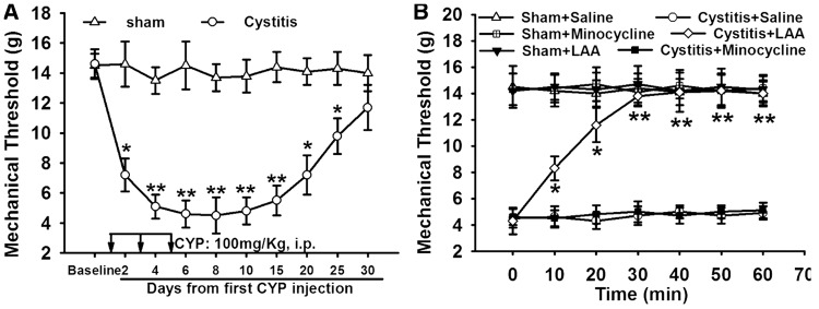
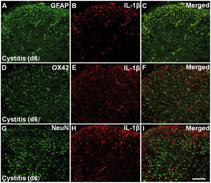
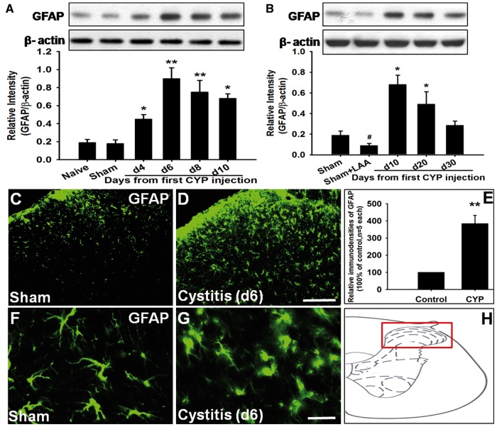
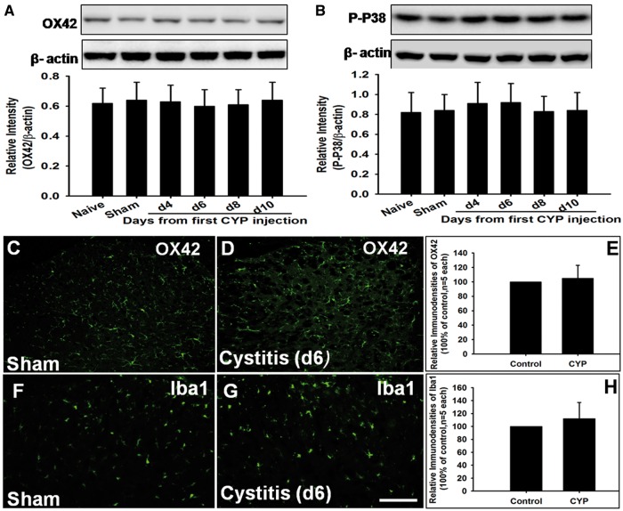
A rat model of bladder pain syndrome/interstitial cystitis was established via systemic injection with cyclophosphamide. Mechanical allodynia was tested with von Frey monofilaments and up-down method. Moreover, Western blots and double immunofluorescence were used to detect the expression and location of glial fibrillary acidic protein, OX42/Iba1, P-P38, NeuN, interleukin (IL)-1β, phosphorylation of N-methyl-D-aspartate receptor 1 (P-NR1), and IL-1 receptor I (IL-1RI) in the L6-S1 spinal cord. We found that glial fibrillary acidic protein rather than OX42/Iba1 or P-P38 was significantly increased in the spinal cord of cyclophosphamide-induced cystitis. L-alpha-aminoadipate but not minocycline markedly attenuated the allodynia. Furthermore, we found that spinal IL-1β was dramatically increased in cyclophosphamide-induced cystitis, and activated astrocytes were the only source of IL-1β release, which contributed to allodynia in cystitis rats. Besides, spinal P-NR1 was statistically increased in cyclophosphamide-induced cystitis and only localized in IL-1RI positive neurons in spinal dorsal horn. Additionally, NR antagonist significantly attenuated the cystitis-induced pain. Interestingly, the time course of the P-NR1 expression paralleled to that of IL-1β or glial fibrillary acidic protein.
Our results demonstrated that astrocytic activation but not microglial activation contributed to the allodynia in cyclophosphamide-induced cystitis and IL-1β released from astrocytes might bind to its endogenous receptor on the neurons inducing the phosphorylation of NR1 subunit, leading to sensory neuronal hyperexcitability and pathological pain.
先前的研究表明,胶质细胞在神经性疼痛的产生和维持中起重要作用。活化的胶质细胞产生多种介质,如促炎细胞因子,这些因子可促进神经元活动和突触可塑性。同样,膀胱疼痛综合征/间质性膀胱炎具有许多神经性疼痛的特征。然而,关于脊髓胶质细胞参与膀胱疼痛综合征/间质性膀胱炎相关病理性疼痛及其潜在机制的相关报道仍然缺乏。本研究调查了膀胱疼痛综合征/间质性膀胱炎大鼠模型中的脊髓胶质细胞活化及潜在分子机制。
通过全身注射环磷酰胺建立膀胱疼痛综合征/间质性膀胱炎大鼠模型。用von Frey细丝和上下法测试机械性异常性疼痛。此外,采用蛋白质免疫印迹法和双重免疫荧光法检测L6-S1脊髓中胶质纤维酸性蛋白、OX42/Iba1、P-P38、NeuN、白细胞介素(IL)-1β、N-甲基-D-天冬氨酸受体1磷酸化(P-NR1)和IL-1受体I(IL-1RI)的表达和定位。我们发现,在环磷酰胺诱导的膀胱炎大鼠脊髓中,胶质纤维酸性蛋白显著增加,而OX42/Iba1或P-P38未增加。L-α-氨基己二酸而非米诺环素可显著减轻异常性疼痛。此外,我们发现环磷酰胺诱导的膀胱炎大鼠脊髓中IL-1β显著增加,活化的星形胶质细胞是IL-1β释放的唯一来源,这导致膀胱炎大鼠出现异常性疼痛。此外,环磷酰胺诱导的膀胱炎大鼠脊髓中P-NR1在统计学上增加,且仅定位于脊髓背角中IL-1RI阳性神经元。此外,NR拮抗剂可显著减轻膀胱炎诱导的疼痛。有趣的是,P-NR1表达的时间进程与IL-1β或胶质纤维酸性蛋白的时间进程平行。
我们的结果表明,星形胶质细胞活化而非小胶质细胞活化导致环磷酰胺诱导的膀胱炎出现异常性疼痛,星形胶质细胞释放的IL-1β可能与其在神经元上的内源性受体结合,诱导NR1亚基磷酸化,导致感觉神经元兴奋性过高和病理性疼痛。