Zhang Rui-Xin, Li Aihui, Liu Bing, Wang Linbo, Ren Ke, Zhang Haiqing, Berman Brian M, Lao Lixing
Center for Integrative Medicine, School of Medicine, University of Maryland, Baltimore, MD 21201, USA Department of Biomedical Sciences, Dental School, University of Maryland, Baltimore, MD 21201, USA.
Pain. 2008 Apr;135(3):232-239. doi: 10.1016/j.pain.2007.05.023. Epub 2007 Aug 6.
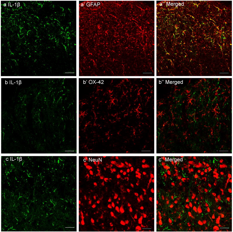
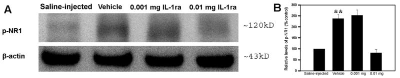
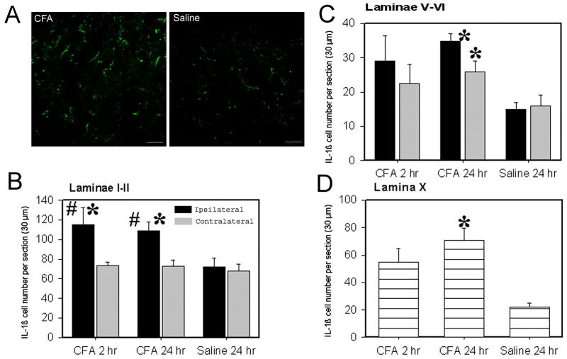
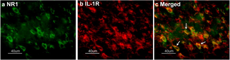
Although it has been shown that pro-inflammatory cytokines such as interleukin-1beta (IL-1beta) facilitate perception of noxious inputs at the spinal level, the mechanisms have not been understood. This study determined the cell type that produces IL-1beta, the co-localization of IL-1 receptor type I (IL-1RI) and Fos and NR1 in the spinal cord, and the effects of IL-1 receptor antagonist (IL-1ra) on NR1 phosphorylation and hyperalgesia in a rat model of inflammatory pain. Phosphorylation of NR1, an essential subunit of the NMDA receptor (NMDAR), is known to modulate NMDAR activity and facilitate pain. Hyperalgesia was induced by injecting complete Freund's adjuvant (CFA, 0.08ml, 40microg Mycobacterium tuberculosis) into one hind paw of each rat. Paw withdrawal latency (PWL) was tested before CFA (-48h) for baseline and 2 and 24h after CFA to assess hyperalgesia. IL-1ra was given (i.t.) 24h before CFA to block the action of basal IL-1beta and 2h prior to each of two PWL tests to block CFA-induced IL-1beta. Spinal cords were removed for double immunostaining of IL-1beta/neuronal marker and IL-1beta/glial cell markers, IL-1RI/Fos and IL-1RI/NR1, and for Western blot to measure NR1 phosphorylation. The data showed that: (1) astrocytes produce IL-1beta, (2) IL-1RI is localized in Fos- and NR1-immunoreactive neurons within the spinal dorsal horn, and (3) IL-1ra at 0.01mg/rat significantly increased PWL (P<0.05) and inhibited NR1 phosphorylation compared to saline control. The results suggest that spinal IL-1beta is produced by astrocytes and enhances NR1 phosphorylation to facilitate inflammatory pain.
尽管已有研究表明,诸如白细胞介素 - 1β(IL - 1β)等促炎细胞因子可在脊髓水平促进对伤害性刺激的感知,但其机制尚未明确。本研究确定了产生IL - 1β的细胞类型、脊髓中I型白细胞介素 - 1受体(IL - 1RI)与Fos和NR1的共定位情况,以及白细胞介素 - 1受体拮抗剂(IL - 1ra)对炎性疼痛大鼠模型中NR1磷酸化和痛觉过敏的影响。已知NMDA受体(NMDAR)的必需亚基NR1的磷酸化可调节NMDAR活性并加剧疼痛。通过向每只大鼠的一只后爪注射完全弗氏佐剂(CFA,0.08ml,40μg结核分枝杆菌)诱导痛觉过敏。在注射CFA前(-48h)测试爪部缩足潜伏期(PWL)作为基线,并在注射CFA后2小时和24小时进行测试以评估痛觉过敏。在注射CFA前24小时给予IL - 1ra(经椎管内注射)以阻断基础IL - 1β的作用,并在两次PWL测试前2小时给予以阻断CFA诱导的IL - 1β。取出脊髓进行IL - 1β/神经元标志物和IL - 1β/胶质细胞标志物、IL - 1RI/Fos和IL - 1RI/NR1的双重免疫染色,并进行蛋白质印迹法以测量NR1磷酸化。数据显示:(1)星形胶质细胞产生IL - 1β;(2)IL - 1RI定位于脊髓背角内Fos和NR1免疫反应阳性的神经元中;(3)与生理盐水对照组相比,0.01mg/大鼠的IL - 1ra显著增加了PWL(P<0.05)并抑制了NR1磷酸化。结果表明,脊髓中的IL - 1β由星形胶质细胞产生,并增强NR1磷酸化以促进炎性疼痛。