Yang Hai-Jing, Xu Wei-Jia, Guan Yi-Hui, Zhang Hui-Wei, Ding Wei-Qun, Rong Lan, Qiu Zhi-Bing, Zhong Liang
Department of Gastroenterology, Huashan Hospital, Fudan University, 12 Wulumuqi Middle Road, Shanghai 200040, China.
PET Center of Huashan Hospital, Fudan University, 518 Wuzhong East Road, Shanghai 200235, China.
Transl Oncol. 2016 Dec;9(6):583-591. doi: 10.1016/j.tranon.2016.08.004.
The purpose of this article is to analyze the expression of Glut-1 and HK-II, the association between their expression and F-FDG accumulation in pancreatic cancer.
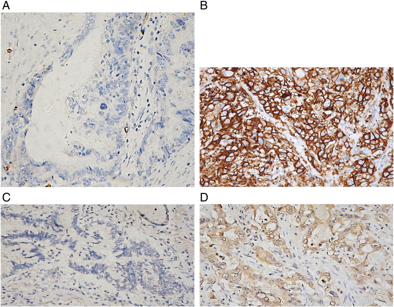
Fifty patients with histologically proven pancreatic cancer were included in this preliminary study, all of whom received F-FDG PET/CT performance before surgery. Immunohistochemical staining of tumor tissue and adjacent normal tissue was performed for Glut-1 and HK-II. By combining proportions and intensity of immunochemical staining, we obtained the modified immunohistological scores for Glut-1 and HK-II respectively. The relationship between expression of Glut-1, HK-II and series of parameters was analyzed, i.e. clinicopathological characteristics, prognosis of patients and SUV of PET-CT.
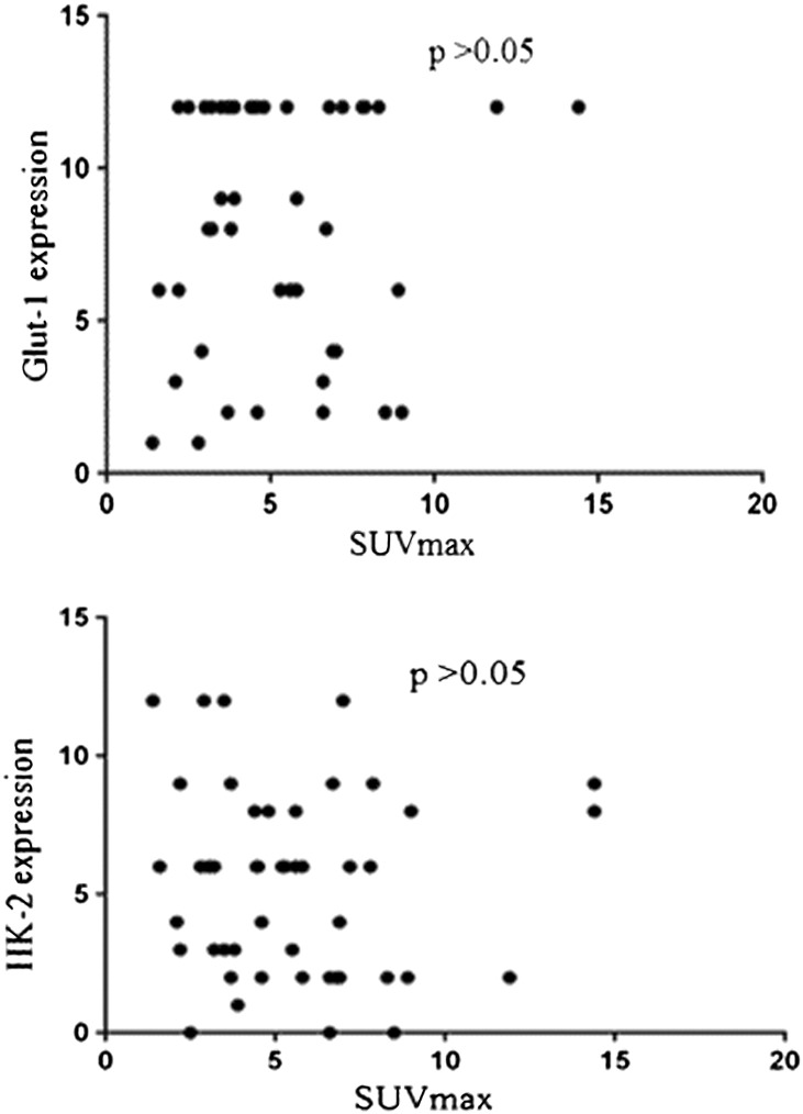
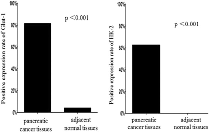
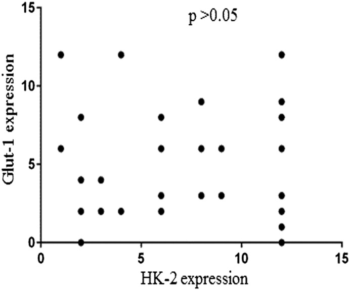
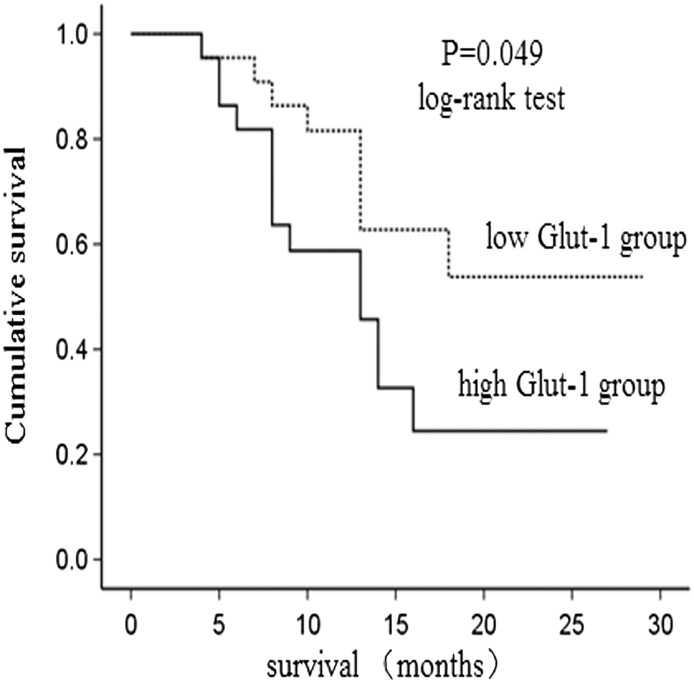
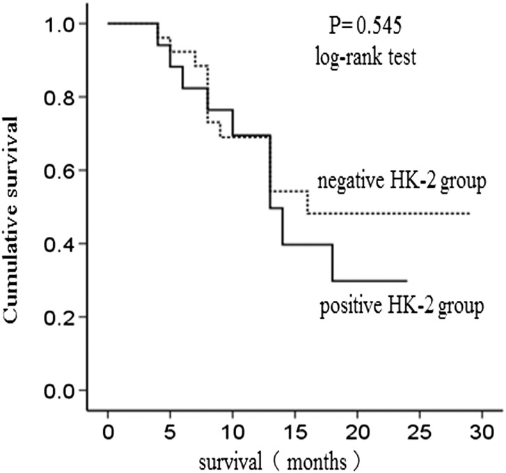
Compared with normal tissue, the Glut-1 and HK-II expression in pancreatic cancer tissue was significantly increased (P<.001). There was no correlation between expression of Glut-1, HK-II and age, gender, tumor size, tumor location, tumor histological type, tumor differentiation, the nerve infiltration, vascular invasion, local infiltration, lymph node metastasis or tumor staging in pancreatic cancer (P>.05). During the follow-up period, the survival curves of low Glut-1 group and high Glut-1 group were statistically different (P=.049). Multivariate analysis (Cox regression) revealed that Glut-1 expression was not associated with mortality (P>.05). No statistical difference was found in the survival curves of negative HK-II group and positive HK-II group (P=.545). There was no correlation between F-FDG uptake and expression of Glut-1 and HK-II(P>.05).
The Glut-1 and HK-II expression in pancreatic cancer tissue was significantly increased. There was no correlation between expression of Glut-1, HK-II and clinicopathological characteristics, prognosis and F-FDG uptake.
本文旨在分析葡萄糖转运蛋白1(Glut-1)和己糖激酶-II(HK-II)的表达情况,以及它们的表达与胰腺癌中氟代脱氧葡萄糖(F-FDG)摄取之间的关联。
本初步研究纳入了50例经组织学证实的胰腺癌患者,所有患者在手术前均接受了F-FDG正电子发射断层扫描/计算机断层扫描(PET/CT)检查。对肿瘤组织和相邻正常组织进行Glut-1和HK-II的免疫组织化学染色。通过结合免疫化学染色的比例和强度,我们分别获得了Glut-1和HK-II的改良免疫组织学评分。分析了Glut-1、HK-II的表达与一系列参数之间的关系,即临床病理特征、患者预后以及PET-CT的标准化摄取值(SUV)。
与正常组织相比,胰腺癌组织中Glut-1和HK-II的表达显著增加(P<0.001)。Glut-1、HK-II的表达与胰腺癌患者的年龄、性别、肿瘤大小、肿瘤位置、肿瘤组织学类型、肿瘤分化程度、神经浸润、血管侵犯、局部浸润、淋巴结转移或肿瘤分期之间均无相关性(P>0.05)。在随访期间,低Glut-1组和高Glut-1组的生存曲线具有统计学差异(P=0.049)。多因素分析(Cox回归)显示,Glut-1表达与死亡率无关(P>0.05)。HK-II阴性组和阳性组的生存曲线无统计学差异(P=0.545)。F-FDG摄取与Glut-1和HK-II的表达之间无相关性(P>0.05)。
胰腺癌组织中Glut-1和HK-II的表达显著增加。Glut-1、HK-II的表达与临床病理特征、预后及F-FDG摄取之间均无相关性。