• 文献检索
  • 文档翻译
  • 深度研究
  • 学术资讯
  • Suppr Zotero 插件Zotero 插件
  • 邀请有礼
  • 套餐&价格
  • 历史记录
应用&插件
Suppr Zotero 插件Zotero 插件浏览器插件Mac 客户端Windows 客户端微信小程序
定价
高级版会员购买积分包购买API积分包
服务
文献检索文档翻译深度研究API 文档MCP 服务
关于我们
关于 Suppr公司介绍联系我们用户协议隐私条款
关注我们

Suppr 超能文献

核心技术专利:CN118964589B侵权必究
粤ICP备2023148730 号-1Suppr @ 2026

文献检索

告别复杂PubMed语法,用中文像聊天一样搜索,搜遍4000万医学文献。AI智能推荐,让科研检索更轻松。

立即免费搜索

文件翻译

保留排版,准确专业,支持PDF/Word/PPT等文件格式,支持 12+语言互译。

免费翻译文档

深度研究

AI帮你快速写综述,25分钟生成高质量综述,智能提取关键信息,辅助科研写作。

立即免费体验

对比增强CT图像上肝脏容积测量的比较:一种半自动方法和两种自动方法

Comparison of liver volumetry on contrast-enhanced CT images: one semiautomatic and two automatic approaches.

作者信息

Cai Wei, He Baochun, Fan Yingfang, Fang Chihua, Jia Fucang

机构信息

Zhujiang Hospital, Southern Medical University; Shenzhen Institutes of Advanced Technology, Chinese Academy of Sciences.

出版信息

J Appl Clin Med Phys. 2016 Nov 8;17(6):118-127. doi: 10.1120/jacmp.v17i6.6485.

DOI:10.1120/jacmp.v17i6.6485
PMID:27929487
原文链接:https://pmc.ncbi.nlm.nih.gov/articles/PMC5690519/
Abstract

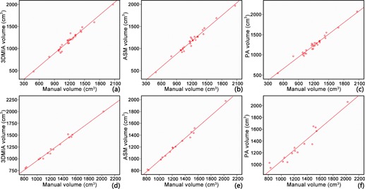
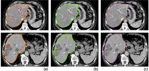
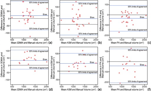
This study was to evaluate the accuracy, consistency, and efficiency of three liver volumetry methods- one interactive method, an in-house-developed 3D medical Image Analysis (3DMIA) system, one automatic active shape model (ASM)-based segmentation, and one automatic probabilistic atlas (PA)-guided segmentation method on clinical contrast-enhanced CT images. Forty-two datasets, including 27 normal liver and 15 space-occupying liver lesion patients, were retrospectively included in this study. The three methods - one semiautomatic 3DMIA, one automatic ASM-based, and one automatic PA-based liver volumetry - achieved an accuracy with VD (volume difference) of -1.69%, -2.75%, and 3.06% in the normal group, respectively, and with VD of -3.20%, -3.35%, and 4.14% in the space-occupying lesion group, respectively. However, the three methods achieved an efficiency of 27.63 mins, 1.26 mins, 1.18 mins on average, respectively, compared with the manual volumetry, which took 43.98 mins. The high intraclass correlation coefficient between the three methods and the manual method indicated an excel-lent agreement on liver volumetry. Significant differences in segmentation time were observed between the three methods (3DMIA, ASM, and PA) and the manual volumetry (p < 0.001), as well as between the automatic volumetries (ASM and PA) and the semiautomatic volumetry (3DMIA) (p < 0.001). The semiautomatic interactive 3DMIA, automatic ASM-based, and automatic PA-based liver volum-etry agreed well with manual gold standard in both the normal liver group and the space-occupying lesion group. The ASM- and PA-based automatic segmentation have better efficiency in clinical use.

摘要

本研究旨在评估三种肝脏容积测量方法的准确性、一致性和效率,这三种方法分别是一种交互式方法、一种自主研发的三维医学图像分析(3DMIA)系统、一种基于主动形状模型(ASM)的自动分割方法以及一种基于概率图谱(PA)的自动分割方法,用于临床对比增强CT图像。本研究回顾性纳入了42个数据集,包括27例正常肝脏患者和15例肝脏占位性病变患者。三种方法,即一种半自动3DMIA、一种基于ASM的自动方法和一种基于PA的自动肝脏容积测量方法,在正常组中的体积差异(VD)准确性分别为-1.69%、-2.75%和3.06%,在占位性病变组中的VD分别为-3.20%、-3.35%和4.14%。然而,与耗时43.98分钟的手工容积测量相比,这三种方法的平均效率分别为27.63分钟、1.26分钟和1.18分钟。三种方法与手工方法之间的组内相关系数较高,表明在肝脏容积测量方面具有良好的一致性。三种方法(3DMIA、ASM和PA)与手工容积测量之间在分割时间上存在显著差异(p<0.001),自动容积测量方法(ASM和PA)与半自动容积测量方法(3DMIA)之间也存在显著差异(p<0.001)。半自动交互式3DMIA、基于ASM的自动方法和基于PA的自动肝脏容积测量在正常肝脏组和占位性病变组中均与手工金标准具有良好的一致性。基于ASM和PA的自动分割在临床应用中具有更高的效率。

https://cdn.ncbi.nlm.nih.gov/pmc/blobs/593b/5690519/b06bf5b1c857/ACM2-17-118-g003.jpg
https://cdn.ncbi.nlm.nih.gov/pmc/blobs/593b/5690519/9345d4a2b402/ACM2-17-118-g001.jpg
https://cdn.ncbi.nlm.nih.gov/pmc/blobs/593b/5690519/e7bdc1deea29/ACM2-17-118-g002.jpg
https://cdn.ncbi.nlm.nih.gov/pmc/blobs/593b/5690519/b06bf5b1c857/ACM2-17-118-g003.jpg
https://cdn.ncbi.nlm.nih.gov/pmc/blobs/593b/5690519/9345d4a2b402/ACM2-17-118-g001.jpg
https://cdn.ncbi.nlm.nih.gov/pmc/blobs/593b/5690519/e7bdc1deea29/ACM2-17-118-g002.jpg
https://cdn.ncbi.nlm.nih.gov/pmc/blobs/593b/5690519/b06bf5b1c857/ACM2-17-118-g003.jpg

相似文献

1
Comparison of liver volumetry on contrast-enhanced CT images: one semiautomatic and two automatic approaches.对比增强CT图像上肝脏容积测量的比较:一种半自动方法和两种自动方法
J Appl Clin Med Phys. 2016 Nov 8;17(6):118-127. doi: 10.1120/jacmp.v17i6.6485.
2
Dr. Liver: A preoperative planning system of liver graft volumetry for living donor liver transplantation.李博士:活体肝移植供肝体积测量的术前规划系统。
Comput Methods Programs Biomed. 2018 May;158:11-19. doi: 10.1016/j.cmpb.2018.01.024. Epub 2018 Jan 31.
3
Shape-intensity prior level set combining probabilistic atlas and probability map constrains for automatic liver segmentation from abdominal CT images.基于形状-强度先验水平集的概率图谱和概率图约束的自动肝脏 CT 图像分割方法。
Int J Comput Assist Radiol Surg. 2016 May;11(5):817-26. doi: 10.1007/s11548-015-1332-9. Epub 2015 Dec 8.
4
A comparative study of automatic image segmentation algorithms for target tracking in MR-IGRT.用于磁共振图像引导放射治疗(MR-IGRT)中目标跟踪的自动图像分割算法的比较研究。
J Appl Clin Med Phys. 2016 Mar 8;17(2):441-460. doi: 10.1120/jacmp.v17i2.5820.
5
Fully automated whole-liver volume quantification on CT-image data: Comparison with manual volumetry using enhanced and unenhanced images as well as two different radiation dose levels and two reconstruction kernels.基于 CT 图像数据的全自动全肝体积定量:增强及非增强图像、两种不同辐射剂量水平和两种重建核的手动容积测量比较。
PLoS One. 2021 Aug 2;16(8):e0255374. doi: 10.1371/journal.pone.0255374. eCollection 2021.
6
Fast automatic 3D liver segmentation based on a three-level AdaBoost-guided active shape model.基于三级AdaBoost引导的主动形状模型的快速自动3D肝脏分割
Med Phys. 2016 May;43(5):2421. doi: 10.1118/1.4946817.
7
Quantitative radiology: automated CT liver volumetry compared with interactive volumetry and manual volumetry.定量放射学:自动 CT 肝脏容积测量与交互式容积测量和手动容积测量比较。
AJR Am J Roentgenol. 2011 Oct;197(4):W706-12. doi: 10.2214/AJR.10.5958.
8
Fully automatic scheme for measuring liver volume in 3D MR images.用于在3D磁共振图像中测量肝脏体积的全自动方案。
Biomed Mater Eng. 2015;26 Suppl 1:S1361-9. doi: 10.3233/BME-151434.
9
Phantom validation of coregistration of PET and CT for image-guided radiotherapy.用于图像引导放射治疗的PET与CT配准的体模验证
Med Phys. 2004 May;31(5):1083-92. doi: 10.1118/1.1688041.
10
Generation of abdominal synthetic CTs from 0.35T MR images using generative adversarial networks for MR-only liver radiotherapy.利用生成对抗网络从 0.35TMR 图像生成腹部合成 CT 用于仅 MR 肝脏放射治疗。
Biomed Phys Eng Express. 2020 Jan 30;6(1):015033. doi: 10.1088/2057-1976/ab6e1f.

引用本文的文献

1
Evaluation of artificial-intelligence-based liver segmentation and its application for longitudinal liver volume measurement.基于人工智能的肝脏分割评估及其在肝脏体积纵向测量中的应用。
Abdom Radiol (NY). 2025 Jun 10. doi: 10.1007/s00261-025-05050-3.
2
Liver volumetry improves evaluation of treatment response to hepatic artery infusion chemotherapy in uveal melanoma patients with liver metastases.肝体积测量可改善转移性葡萄膜黑色素瘤患者肝动脉化疗栓塞治疗反应的评估。
Radiol Oncol. 2024 Nov 28;58(4):509-516. doi: 10.2478/raon-2024-0063. eCollection 2024 Dec 1.
3
Diagnostic accuracy of 3D imaging combined with intra-operative ultrasound in the prediction of post-hepatectomy liver failure.

本文引用的文献

1
Fast automatic 3D liver segmentation based on a three-level AdaBoost-guided active shape model.基于三级AdaBoost引导的主动形状模型的快速自动3D肝脏分割
Med Phys. 2016 May;43(5):2421. doi: 10.1118/1.4946817.
2
Impact of three-dimensional reconstruction technique in the operation planning of centrally located hepatocellular carcinoma.三维重建技术在肝门部肝细胞癌手术规划中的应用
J Am Coll Surg. 2015 Jan;220(1):28-37. doi: 10.1016/j.jamcollsurg.2014.09.023. Epub 2014 Oct 15.
3
Accuracy of preoperative automatic measurement of the liver volume by CT-scan combined to a 3D virtual surgical planning software (3DVSP).
三维成像联合术中超声在预测肝切除术后肝衰竭中的诊断准确性
J Gastrointest Oncol. 2022 Jun;13(3):1224-1236. doi: 10.21037/jgo-22-282.
4
Deep Learning CT-based Quantitative Visualization Tool for Liver Volume Estimation: Defining Normal and Hepatomegaly.深度学习 CT 定量可视化工具用于肝体积估计:定义正常肝和肝肿大。
Radiology. 2022 Feb;302(2):336-342. doi: 10.1148/radiol.2021210531. Epub 2021 Oct 26.
5
Fully automated whole-liver volume quantification on CT-image data: Comparison with manual volumetry using enhanced and unenhanced images as well as two different radiation dose levels and two reconstruction kernels.基于 CT 图像数据的全自动全肝体积定量:增强及非增强图像、两种不同辐射剂量水平和两种重建核的手动容积测量比较。
PLoS One. 2021 Aug 2;16(8):e0255374. doi: 10.1371/journal.pone.0255374. eCollection 2021.
6
Atlas-based liver segmentation for nonhuman primate research.基于图谱的非人类灵长类动物肝脏分割。
Int J Comput Assist Radiol Surg. 2020 Oct;15(10):1631-1638. doi: 10.1007/s11548-020-02225-9. Epub 2020 Jul 9.
7
Automated Segmentation of Tissues Using CT and MRI: A Systematic Review.基于 CT 和 MRI 的组织自动分割:系统评价。
Acad Radiol. 2019 Dec;26(12):1695-1706. doi: 10.1016/j.acra.2019.07.006. Epub 2019 Aug 10.
8
Inter-observer variability of manual contour delineation of structures in CT.CT 中手动勾画结构轮廓的观察者间变异性。
Eur Radiol. 2019 Mar;29(3):1391-1399. doi: 10.1007/s00330-018-5695-5. Epub 2018 Sep 7.
通过CT扫描结合三维虚拟手术规划软件(3DVSP)对肝脏体积进行术前自动测量的准确性。
Surg Endosc. 2014 Dec;28(12):3408-12. doi: 10.1007/s00464-014-3611-x. Epub 2014 Jun 14.
4
Liver planning software accurately predicts postoperative liver volume and measures early regeneration.肝脏规划软件能准确预测术后肝脏体积并测量早期肝脏再生情况。
J Am Coll Surg. 2014 Aug;219(2):199-207. doi: 10.1016/j.jamcollsurg.2014.02.027. Epub 2014 Mar 27.
5
Multi-organ segmentation based on spatially-divided probabilistic atlas from 3D abdominal CT images.基于3D腹部CT图像空间划分概率图谱的多器官分割
Med Image Comput Comput Assist Interv. 2013;16(Pt 2):165-72. doi: 10.1007/978-3-642-40763-5_21.
6
Computerized liver volumetry on MRI by using 3D geodesic active contour segmentation.基于 3D 测地线主动轮廓分割的 MRI 计算机化肝脏体积测量。
AJR Am J Roentgenol. 2014 Jan;202(1):152-9. doi: 10.2214/AJR.13.10812.
7
Liver segmentation based on Snakes Model and improved GrowCut algorithm in abdominal CT image.基于 Snake 模型和改进的 GrowCut 算法的腹部 CT 图像肝脏分割。
Comput Math Methods Med. 2013;2013:958398. doi: 10.1155/2013/958398. Epub 2013 Aug 26.
8
Outcomes of hepatectomy for hepatolithiasis based on 3-dimensional reconstruction technique.基于三维重建技术的肝切除术治疗肝内胆管结石的疗效。
J Am Coll Surg. 2013 Aug;217(2):280-8. doi: 10.1016/j.jamcollsurg.2013.03.017.
9
Sample size formulas for estimating intraclass correlation coefficients with precision and assurance.估计具有精度和保证的组内相关系数的样本量公式。
Stat Med. 2012 Dec 20;31(29):3972-81. doi: 10.1002/sim.5466. Epub 2012 Jul 4.
10
Assessing hepatomegaly: automated volumetric analysis of the liver.评估肝肿大:肝脏的自动容积分析。
Acad Radiol. 2012 May;19(5):588-98. doi: 10.1016/j.acra.2012.01.015. Epub 2012 Feb 22.