Sprenger Julia U, Bork Nadja I, Herting Jonas, Fischer Thomas H, Nikolaev Viacheslav O
Institute of Experimental Cardiovascular Research, University Medical Center Hamburg-Eppendorf, Hamburg, Germany.
Clinic of Cardiology and Pulmonology, Heart Research Center Göttingen, University Medical Center Göttingen, Göttingen, Germany.
PLoS One. 2016 Dec 8;11(12):e0167974. doi: 10.1371/journal.pone.0167974. eCollection 2016.
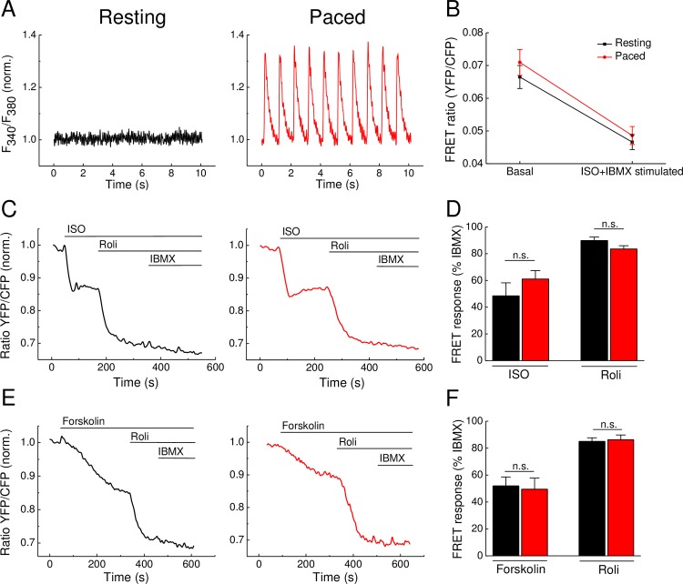
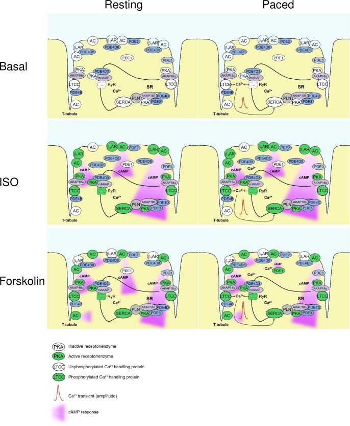
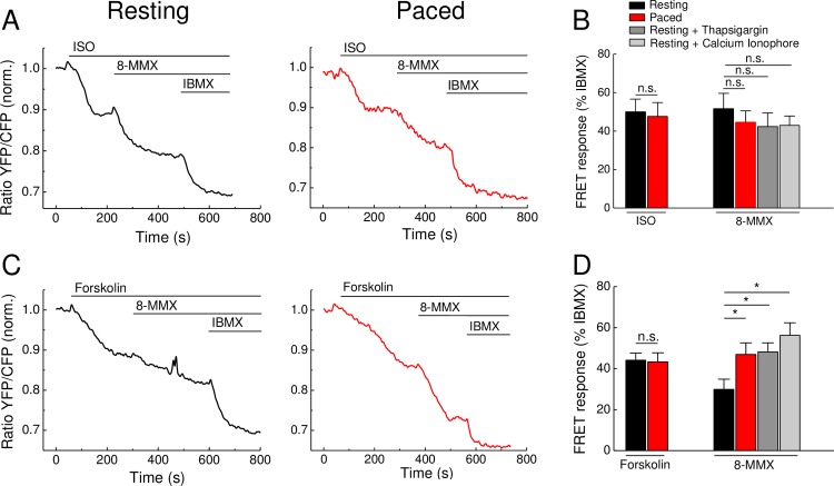
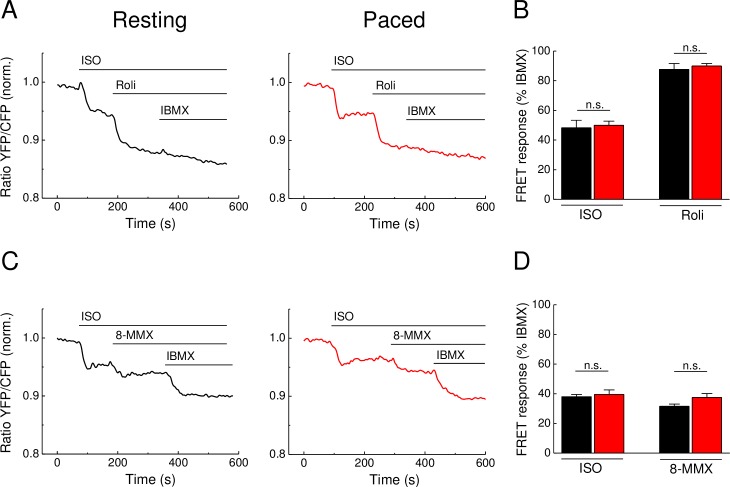
Calcium (Ca2+) and 3',5'-cyclic adenosine monophosphate (cAMP) play a critical role for cardiac excitation-contraction-coupling. Both second messengers are known to interact with each other, for example via Ca2+-dependent modulation of phosphodiesterase 1 (PDE1) and adenylyl cyclase 5/6 (AC 5/6) activities, which is supposed to occur especially at the local level in distinct subcellular microdomains. Currently, many studies analyze global and local cAMP signaling and its regulation in resting cardiomyocytes devoid of electrical stimulation. For example, Förster resonance energy transfer (FRET) microscopy is a popular approach for visualization of real time cAMP dynamics performed in resting cardiomyocytes to avoid potential contractility-related movement artifacts. However, it is unknown whether such data are comparable with the cell behavior under more physiologically relevant conditions during contraction. Here, we directly compare the cAMP-FRET responses to AC stimulation and PDE inhibition in resting vs. paced adult mouse ventricular cardiomyocytes for both cytosolic and subsarcolemmal microdomains. Interestingly, no significant differences in cAMP dynamics could be detected after β-adrenergic (isoproterenol) stimulation, suggesting low impact of rapidly changing contractile Ca2+ concentrations on cytosolic cAMP levels associated with AC activation. However, the contribution of the calcium-dependent PDE1, but not of the Ca2+-insensitive PDE4, to the regulation of cAMP levels after forskolin stimulation was significantly increased. This increase could be mimicked by pretreatment of resting cells with Ca2+ elevating agents. Ca2+ imaging demonstrated significantly higher amplitudes of Ca2+ transients in forskolin than in isoproterenol stimulated cells, suggesting that forskolin stimulation might lead to stronger activation of PDE1. In conclusion, changes in intracellular Ca2+ during cardiomyocyte contraction dynamically interact with cAMP levels, especially after strong AC stimulation. The use of resting cells for FRET-based measurements of cAMP can be justified under β-adrenergic stimulation, while the reliable analysis of PDE1 effects may require electric field stimulation.
钙(Ca2+)和3',5'-环磷酸腺苷(cAMP)在心脏兴奋-收缩偶联中起关键作用。已知这两种第二信使会相互作用,例如通过Ca2+依赖性调节磷酸二酯酶1(PDE1)和腺苷酸环化酶5/6(AC 5/6)的活性,这种相互作用被认为尤其发生在不同亚细胞微区的局部水平。目前,许多研究分析了静息心肌细胞中全局和局部的cAMP信号传导及其调节,这些细胞没有电刺激。例如,荧光共振能量转移(FRET)显微镜是一种用于可视化静息心肌细胞中实时cAMP动态变化的常用方法,以避免潜在的与收缩性相关的运动伪影。然而,尚不清楚这些数据是否与收缩过程中更生理相关条件下的细胞行为具有可比性。在这里,我们直接比较了静息和起搏的成年小鼠心室心肌细胞中胞质和肌膜下微区对AC刺激和PDE抑制的cAMP-FRET反应。有趣的是,在β-肾上腺素能(异丙肾上腺素)刺激后,未检测到cAMP动态变化的显著差异,这表明快速变化的收缩性Ca2+浓度对与AC激活相关的胞质cAMP水平影响较小。然而,钙依赖性PDE1而非Ca2+不敏感的PDE4对福斯高林刺激后cAMP水平调节的贡献显著增加。用Ca2+升高剂预处理静息细胞可模拟这种增加。Ca2+成像显示,福斯高林刺激的细胞中Ca2+瞬变的幅度明显高于异丙肾上腺素刺激的细胞,这表明福斯高林刺激可能导致PDE1更强的激活。总之,心肌细胞收缩过程中细胞内Ca2+的变化与cAMP水平动态相互作用,尤其是在强烈的AC刺激后。在β-肾上腺素能刺激下,使用静息细胞进行基于FRET的cAMP测量是合理的,而对PDE1效应的可靠分析可能需要电场刺激。