Aparicio Javier, Carreño Mar, Bargalló Núria, Setoain Xavier, Rubí Sebastià, Rumià Jordi, Falcón Carles, Calvo Anna, Martí-Fuster Berta, Padilla Nelly, Boget Teresa, Pintor Luís, Donaire Antonio
Hospital Clínic, Epilepsy Program, Department of Neurology, Neuroscience Institute, CP 08036, Barcelona, Spain.
Hospital Clínic, Epilepsy Program, Department of Neurology, Neuroscience Institute, CP 08036, Barcelona, Spain; Institut d'investigacions Biomèdiques August Pi i Sunyer (IDIBAPS), CP 08036, Barcelona, Spain.
Neuroimage Clin. 2016 May 6;12:976-989. doi: 10.1016/j.nicl.2016.05.002. eCollection 2016.
Several studies using F-fluorodeoxyglucose positron emission tomography (F-FDG-PET) or diffusion tensor imaging (DTI) have found both temporal and extratemporal abnormalities in patients with mesial temporal lobe epilepsy with ipsilateral hippocampal sclerosis (MTLE-HS), but data are lacking about the findings of both techniques in the same patients. We aimed to determine whether the extent of F-FDG-PET hypometabolism is related to DTI abnormalities.
Twenty-one patients with MTLE-HS underwent comprehensive preoperative evaluation; 18 (86%) of these underwent epilepsy surgery. We analyzed and compared the pattern of white matter (WM) alterations on DTI and cortical hypometabolism on F-FDG-PET.
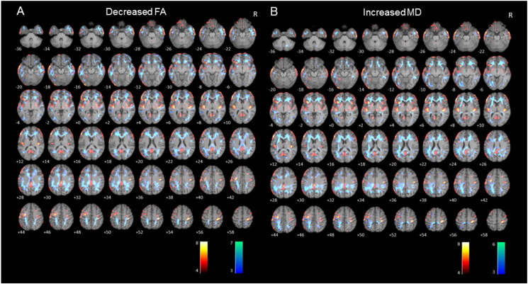
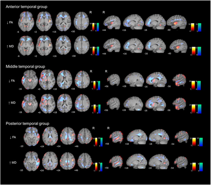
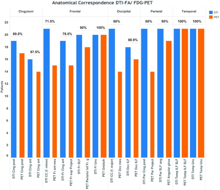
We found widespread temporal and extratemporal F-FDG-PET and DTI abnormalities. Patterns of WM abnormalities and cortical glucose hypometabolism involved similar brain regions, being more extensive in the left than the right MTLE-HS. We classified patients into three groups according to temporal F-FDG-PET patterns: hypometabolism restricted to the anterior third (n = 7), hypometabolism extending to the middle third (n = 7), and hypometabolism extending to the posterior third (n = 7). Patients with anterior temporal hypometabolism showed DTI abnormalities in anterior association and commissural tracts while patients with posterior hypometabolism showed WM alterations in anterior and posterior tracts.
Patients with MTLE-HS have widespread metabolic and microstructural abnormalities that involve similar regions. The distribution patterns of these gray and white matter abnormalities differ between patients with left or right MTLE, but also with the extent of the F-FDG-PET hypometabolism along the epileptogenic temporal lobe. These findings suggest a variable network involvement among patients with MTLE-HS.
多项使用氟脱氧葡萄糖正电子发射断层扫描(F-FDG-PET)或弥散张量成像(DTI)的研究发现,内侧颞叶癫痫伴同侧海马硬化(MTLE-HS)患者存在颞叶及颞叶外异常,但缺乏同一患者这两种技术检查结果的相关数据。我们旨在确定F-FDG-PET代谢减低的程度是否与DTI异常有关。
21例MTLE-HS患者接受了全面的术前评估;其中18例(86%)接受了癫痫手术。我们分析并比较了DTI上白质(WM)改变模式与F-FDG-PET上皮质代谢减低情况。
我们发现广泛的颞叶及颞叶外F-FDG-PET和DTI异常。WM异常模式和皮质葡萄糖代谢减低涉及相似的脑区,在左侧MTLE-HS中比右侧更广泛。我们根据颞叶F-FDG-PET模式将患者分为三组:代谢减低局限于前三分之一(n = 7)、代谢减低延伸至中三分之一(n = 7)、代谢减低延伸至后三分之一(n = 7)。颞叶前部代谢减低的患者在前联合和连合束中显示DTI异常,而后部代谢减低的患者在前束和后束中显示WM改变。
MTLE-HS患者存在广泛的代谢和微观结构异常,累及相似区域。这些灰质和白质异常的分布模式在左侧或右侧MTLE患者之间存在差异,而且还与致痫颞叶上F-FDG-PET代谢减低的程度有关。这些发现提示MTLE-HS患者中存在可变的网络受累情况。