Nakamura Hiroki, Kudo Shin-Ei, Misawa Masashi, Kataoka Shinichi, Wakamura Kunihiko, Hayashi Takemasa, Kudo Toyoki, Mori Yuichi, Takeda Kenichi, Ichimasa Katsuro, Miyachi Hideyuki, Katagiri Atushi, Ishida Fumio, Inoue Haruhiro
Digestive Disease Center, Showa University, Northern Yokohama Hospital, Yokohama, Japan.
Digestive Disease Center, Showa University, Koto-Toyosu Hospital, Tokyo, Japan.
Endosc Int Open. 2016 Dec;4(12):E1280-E1285. doi: 10.1055/s-0042-117629. Epub 2016 Nov 10.
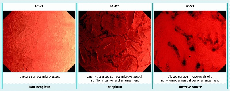
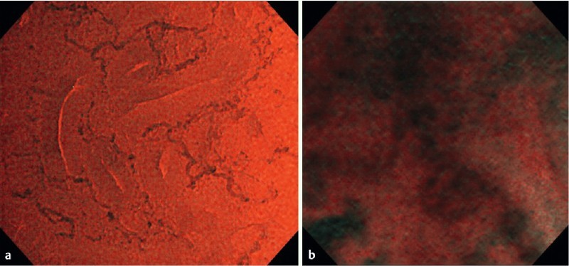
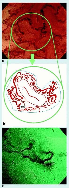
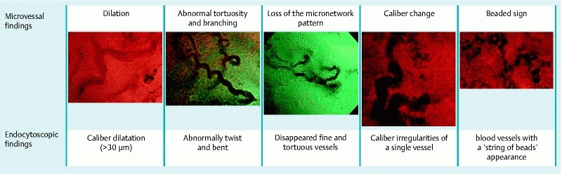
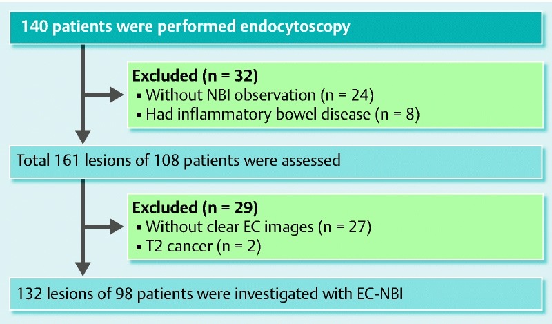
Magnifying narrow-band imaging (NBI) is useful for examination of colorectal lesions, and endocytoscopy (EC) allows diagnostic evaluation of structural atypia, nuclear atypia, and vascular structures of colorectal tumors. The aim of this study was to examine surface microvessels in deep invasive colorectal cancer using EC with a new NBI video processor system. We retrospectively assessed 132 colorectal neoplastic lesions: 81 adenomas, 18 intramucosal cancers, 4 submucosal slightly invasive cancers, and 29 submucosal deep invasive cancers. Detailed vascular findings commonly seen in submucosal deep invasive carcinomas included > 2-fold vasodilatation seen in adenomas, abnormal tortuosity and branching, loss of the micro-network pattern, caliber change in > 2 places in a single blood vessel, and blood vessels not visible in a line because they appear like a string of beads (beaded sign). Univariate analysis revealed 4 vascular findings that were strongly predictive of submucosal deep invasion: vasodilatation (odds ratio [OR] 9.31; 95 % confidence interval [CI] 3.57 - 24.30), loss of the micro-network pattern (OR 61.60; 95 % CI 17.87 - 212.29), caliber change (OR 35.7; 95 % CI 9.16 - 139.14), and the beaded sign (OR 45.90; 95 % CI 5.50 - 382.73). Detailed assessment of ultra-magnified microvessels could improve the diagnostic performance for submucosal deep invasive cancer.
UMIN-CTR000014033.
放大窄带成像(NBI)对结直肠病变检查有用,而内镜下活检(EC)可对结直肠肿瘤的结构异型性、核异型性和血管结构进行诊断评估。本研究的目的是使用配备新型NBI视频处理器系统的EC检查深部浸润性结直肠癌的表面微血管。我们回顾性评估了132例结直肠肿瘤性病变:81例腺瘤、18例黏膜内癌、4例黏膜下微浸润癌和29例黏膜下深部浸润癌。黏膜下深部浸润癌中常见的详细血管表现包括:腺瘤中可见的血管扩张>2倍、异常迂曲和分支、微血管网络模式消失、单个血管中>2处管径改变以及血管呈串珠样排列(串珠征)而无法连成一线。单因素分析显示,4种血管表现强烈提示黏膜下深部浸润:血管扩张(比值比[OR]9.31;95%置信区间[CI]3.57 - 24.30)、微血管网络模式消失(OR 61.60;95% CI 17.87 - 212.29)、管径改变(OR 35.7;95% CI 9.16 - 139.14)和串珠征(OR 45.90;95% CI 5.50 - 382.73)。对超放大微血管的详细评估可提高黏膜下深部浸润癌的诊断性能。
UMIN - CTR000014033。