• 文献检索
  • 文档翻译
  • 深度研究
  • 学术资讯
  • Suppr Zotero 插件Zotero 插件
  • 邀请有礼
  • 套餐&价格
  • 历史记录
应用&插件
Suppr Zotero 插件Zotero 插件浏览器插件Mac 客户端Windows 客户端微信小程序
定价
高级版会员购买积分包购买API积分包
服务
文献检索文档翻译深度研究API 文档MCP 服务
关于我们
关于 Suppr公司介绍联系我们用户协议隐私条款
关注我们

Suppr 超能文献

核心技术专利:CN118964589B侵权必究
粤ICP备2023148730 号-1Suppr @ 2026

文献检索

告别复杂PubMed语法,用中文像聊天一样搜索,搜遍4000万医学文献。AI智能推荐,让科研检索更轻松。

立即免费搜索

文件翻译

保留排版,准确专业,支持PDF/Word/PPT等文件格式,支持 12+语言互译。

免费翻译文档

深度研究

AI帮你快速写综述,25分钟生成高质量综述,智能提取关键信息,辅助科研写作。

立即免费体验

结晶紫和亚甲蓝双重染色适用于结肠内镜检查:一项体内前瞻性初步研究。

Double staining with crystal violet and methylene blue is appropriate for colonic endocytoscopy: an in vivo prospective pilot study.

作者信息

Ichimasa Katsuro, Kudo Shin-ei, Mori Yuichi, Wakamura Kunihiko, Ikehara Nobunao, Kutsukawa Makoto, Takeda Kenichi, Misawa Masashi, Kudo Toyoki, Miyachi Hideyuki, Yamamura Fuyuhiko, Ohkoshi Shogo, Hamatani Shigeharu, Inoue Haruhiro

机构信息

Digestive Disease Center, Showa University Northern Yokohama Hospital, Yokohama, Japan.

出版信息

Dig Endosc. 2014 May;26(3):403-8. doi: 10.1111/den.12164. Epub 2013 Sep 10.

DOI:10.1111/den.12164
PMID:24016362
原文链接:https://pmc.ncbi.nlm.nih.gov/articles/PMC4232925/
Abstract

BACKGROUND AND AIM

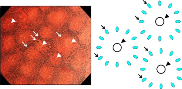
Endocytoscopy (EC) at ultra-high magnification enables in vivo visualization of cellular atypia of gastrointestinal mucosae. Clear images are essential for precise diagnosis by EC. The aim of the present study was to evaluate the optimal staining method for EC in the colon.

METHODS

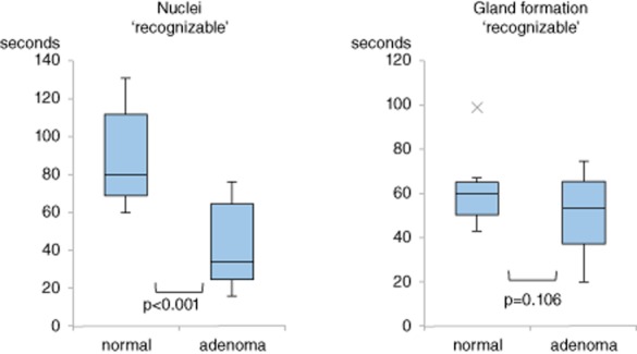
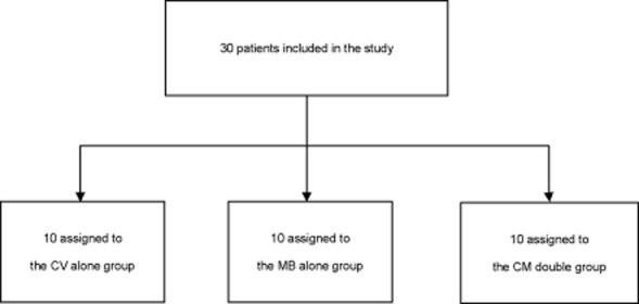
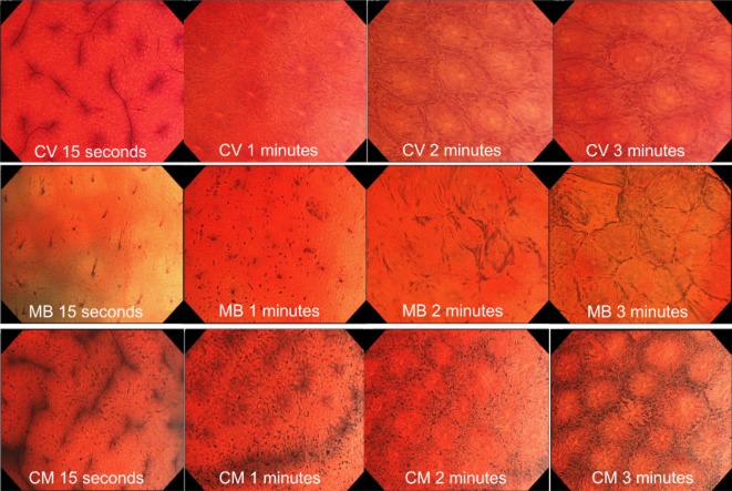
Thirty prospectively enrolled patients were allocated 1:1:1 to three distinct staining methods: 0.05% crystal violet (CV) alone, 1% methylene blue (MB) alone, or CV+MB (CM double). Normal rectal mucosae were stained with each dye and videos of EC images were recorded. Visibility of nuclei and gland formation after staining were evaluated as 'recognizable' or 'not recognizable'. Time for each parameter to become 'recognizable' was measured, and the average times for the three staining regimens were compared.

RESULTS

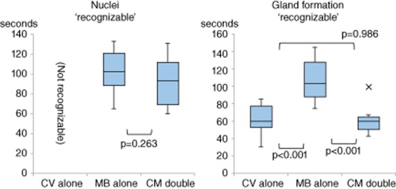
MB alone and CM double staining resulted in 'recognizable' (102 ± 27 vs 89 ± 22 s, P=0.263) nuclei within comparable periods of time, whereas CV alone was unable to identify nuclei. Gland formation became 'recognizable' sooner after CM double staining than after MB alone (61 ± 16 vs 108 ± 24 s, P<0.001).

CONCLUSIONS

Double staining with CV and MB, which rapidly provided recognizable images of both nuclei and gland formation, is an appropriate staining regimen for colonic EC.

摘要

背景与目的

超高倍放大的内镜检查术(EC)能够在体内观察胃肠道黏膜的细胞异型性。清晰的图像对于EC的精确诊断至关重要。本研究的目的是评估结肠EC的最佳染色方法。

方法

将30例前瞻性纳入的患者按1:1:1分配至三种不同的染色方法:单独使用0.05%结晶紫(CV)、单独使用1%亚甲蓝(MB)或CV+MB(CM双重染色)。用每种染料对正常直肠黏膜进行染色,并记录EC图像的视频。将染色后细胞核和腺体形成的可见性评估为“可识别”或“不可识别”。测量每个参数达到“可识别”的时间,并比较三种染色方案的平均时间。

结果

单独使用MB和CM双重染色在相当的时间段内使细胞核“可识别”(分别为102±27秒和89±22秒,P=0.263),而单独使用CV无法识别细胞核。CM双重染色后腺体形成比单独使用MB更快“可识别”(分别为61±16秒和108±24秒,P<0.001)。

结论

CV和MB双重染色能快速提供可识别的细胞核和腺体形成图像,是结肠EC合适的染色方案。

https://cdn.ncbi.nlm.nih.gov/pmc/blobs/8b35/4232925/0f9c6ce9199a/den0026-0403-f5.jpg
https://cdn.ncbi.nlm.nih.gov/pmc/blobs/8b35/4232925/63f08166c9b1/den0026-0403-f1.jpg
https://cdn.ncbi.nlm.nih.gov/pmc/blobs/8b35/4232925/a012c3b95671/den0026-0403-f2.jpg
https://cdn.ncbi.nlm.nih.gov/pmc/blobs/8b35/4232925/bb960c486eff/den0026-0403-f3.jpg
https://cdn.ncbi.nlm.nih.gov/pmc/blobs/8b35/4232925/63167784e3df/den0026-0403-f4.jpg
https://cdn.ncbi.nlm.nih.gov/pmc/blobs/8b35/4232925/0f9c6ce9199a/den0026-0403-f5.jpg

相似文献

1
Double staining with crystal violet and methylene blue is appropriate for colonic endocytoscopy: an in vivo prospective pilot study.结晶紫和亚甲蓝双重染色适用于结肠内镜检查:一项体内前瞻性初步研究。
Dig Endosc. 2014 May;26(3):403-8. doi: 10.1111/den.12164. Epub 2013 Sep 10.
2
Ex-vivo study of high-magnification chromoendoscopy in the gastrointestinal tract to determine the optimal staining conditions for endocytoscopy.胃肠道高倍放大色素内镜的体外研究,以确定内镜下活检的最佳染色条件。
Endoscopy. 2006 Nov;38(11):1115-21. doi: 10.1055/s-2006-944915.
3
Endocytoscopy allows accurate in vivo differentiation of mucosal inflammatory cells in IBD: a pilot study.内镜下细胞学法可准确鉴别 IBD 黏膜炎症细胞:一项初步研究。
Inflamm Bowel Dis. 2013 Feb;19(2):356-62. doi: 10.1002/ibd.23025.
4
Dye solution optimizing staining conditions for endocytoscopy for normal villi and superficial epithelial tumors in the duodenum.用于十二指肠正常绒毛和浅表上皮肿瘤的内镜检查染色条件优化的染料溶液。
Ann Gastroenterol. 2019 Jul-Aug;32(4):378-386. doi: 10.20524/aog.2019.0382. Epub 2019 May 10.
5
Endocytoscopy Is Useful for the Diagnosis of Superficial Nonampullary Duodenal Epithelial Tumors.内镜下黏膜切除术治疗胃固有肌层来源黏膜下肿瘤的临床价值
Digestion. 2021;102(6):895-902. doi: 10.1159/000516512. Epub 2021 Jun 7.
6
histological diagnosis for gastric cancer using endocytoscopy.使用内镜下细胞学诊断胃癌。
World J Gastroenterol. 2017 Oct 7;23(37):6894-6901. doi: 10.3748/wjg.v23.i37.6894.
7
Accuracy of diagnosing invasive colorectal cancer using computer-aided endocytoscopy.使用计算机辅助内镜下细胞学检查诊断浸润性结直肠癌的准确性。
Endoscopy. 2017 Aug;49(8):798-802. doi: 10.1055/s-0043-105486. Epub 2017 May 4.
8
Endocytoscopy can provide additional diagnostic ability to magnifying chromoendoscopy for colorectal neoplasms.内镜黏膜下剥离术可以为大肠肿瘤的放大色素内镜检查提供额外的诊断能力。
J Gastroenterol Hepatol. 2014 Jan;29(1):83-90. doi: 10.1111/jgh.12374.
9
[Ultrahigh magnifying endoscopy: development of CM double staining for endocytoscopy and its safety].[超高倍率内镜检查:用于内镜下活检的CM双重染色技术的发展及其安全性]
Nihon Rinsho. 2010 Jul;68(7):1247-52.
10
Endocytoscopy is a promising modality with high diagnostic accuracy for gastric cancer.内镜超声检查是一种对胃癌具有高诊断准确性的有前景的检查方式。
Endoscopy. 2015 Jan;47(1):19-25. doi: 10.1055/s-0034-1377965. Epub 2014 Sep 15.

引用本文的文献

1
Diagnostic performance of endocytoscopy with normal pit-like structure sign for colorectal low-grade adenoma compared with conventional modalities.与传统方法相比,具有正常凹坑样结构征象的内镜下活检对结直肠低级别腺瘤的诊断性能。
DEN Open. 2023 May 8;4(1):e238. doi: 10.1002/deo2.238. eCollection 2024 Apr.
2
Comparison of Ultra-Magnifying Endocytoscopic and Hematoxylin-Eosin-Stained Images of Lung Specimens.肺标本的超放大内镜图像与苏木精-伊红染色图像的比较。
Diagnostics (Basel). 2023 Mar 6;13(5):1003. doi: 10.3390/diagnostics13051003.
3
Future of Endoscopy in Inflammatory Bowel Diseases (IBDs).

本文引用的文献

1
Comprehensive diagnostic ability of endocytoscopy compared with biopsy for colorectal neoplasms: a prospective randomized noninferiority trial.内镜下细胞学诊断与活检对结直肠肿瘤的诊断效能比较:一项前瞻性随机非劣效性试验。
Endoscopy. 2013;45(2):98-105. doi: 10.1055/s-0032-1325932. Epub 2013 Jan 10.
2
Endocytoscopy allows accurate in vivo differentiation of mucosal inflammatory cells in IBD: a pilot study.内镜下细胞学法可准确鉴别 IBD 黏膜炎症细胞:一项初步研究。
Inflamm Bowel Dis. 2013 Feb;19(2):356-62. doi: 10.1002/ibd.23025.
3
Recent advancement of observing living cells in the esophagus using CM double staining: endocytoscopic atypia classification.
炎症性肠病(IBD)内镜检查的未来
Cureus. 2022 Sep 25;14(9):e29567. doi: 10.7759/cureus.29567. eCollection 2022 Sep.
4
Molecular Imaging of Ulex Europaeus Agglutinin in Colorectal Cancer Using Confocal Laser Endomicroscopy (With Video).利用共聚焦激光内镜显微镜对结直肠癌中荆豆凝集素进行分子成像研究(附视频)
Front Oncol. 2021 Dec 20;11:792420. doi: 10.3389/fonc.2021.792420. eCollection 2021.
5
Clinical Efficacy of Endocytoscopy for Gastrointestinal Endoscopy.内镜超声在胃肠内镜检查中的临床疗效
Clin Endosc. 2021 Jul;54(4):455-463. doi: 10.5946/ce.2021.165. Epub 2021 Jul 7.
6
Ultra-magnifying narrow-band imaging for endoscopic diagnosis of gastric intestinal metaplasia: a pilot image analysis study.用于胃肠化生内镜诊断的超放大窄带成像:一项试点图像分析研究
Endosc Int Open. 2021 Apr;9(4):E522-E529. doi: 10.1055/a-1352-2500. Epub 2021 Mar 17.
7
A novel Lugol's iodine staining technique to visualize the upper margin of the surgical anal canal intraoperatively for Hirschsprung disease: a case series.一种新型卢戈氏碘染色技术,可在术中可视化先天性巨结肠症的手术肛门内口上缘:病例系列研究。
BMC Surg. 2020 Dec 4;20(1):317. doi: 10.1186/s12893-020-00986-3.
8
Small invasive colon cancer with adenoma observed by endocytoscopy: A case report.经内镜观察到的伴有腺瘤的小侵袭性结肠癌:病例报告
World J Gastrointest Endosc. 2020 Sep 16;12(9):304-309. doi: 10.4253/wjge.v12.i9.304.
9
Advances in optical gastrointestinal endoscopy: a technical review.光学胃肠道内镜的进展:技术综述。
Mol Oncol. 2021 Oct;15(10):2580-2599. doi: 10.1002/1878-0261.12792. Epub 2020 Sep 19.
10
Endocytoscopy: technology and clinical application in the lower GI tract.内镜超声检查术:在下消化道中的技术与临床应用
Transl Gastroenterol Hepatol. 2020 Jul 5;5:40. doi: 10.21037/tgh.2019.12.04. eCollection 2020.
使用 CM 双重染色观察食管活细胞的最新进展:内视镜下不典型分类。
Dis Esophagus. 2012 Apr;25(3):235-41. doi: 10.1111/j.1442-2050.2011.01241.x. Epub 2011 Sep 2.
4
Diagnosis of colorectal lesions with a novel endocytoscopic classification - a pilot study.新型细胞内镜分类诊断结直肠病变-初步研究。
Endoscopy. 2011 Oct;43(10):869-75. doi: 10.1055/s-0030-1256663. Epub 2011 Aug 11.
5
In vivo visualization of trophozoites in patients with amoebic colitis by using a newly developed endocytoscope.应用新型内视镜观察阿米巴结肠炎患者的滋养体。
Gastrointest Endosc. 2010 Sep;72(3):643-6. doi: 10.1016/j.gie.2010.04.031. Epub 2010 Jun 25.
6
Endocytoscopy can identify dysplasia in aberrant crypt foci of the colorectum: a prospective in vivo study.内镜下活检可识别结直肠异常隐窝病灶中的发育异常:一项前瞻性体内研究。
Endoscopy. 2009 Feb;41(2):129-32. doi: 10.1055/s-0028-1103452. Epub 2009 Feb 12.
7
Can an endocytoscope system (ECS) predict histology in neoplastic lesions?内镜超声系统(ECS)能否预测肿瘤性病变的组织学特征?
Endoscopy. 2007 Jun;39(6):497-501. doi: 10.1055/s-2007-966446.
8
Detection of tissue abnormalities in normal mucosa surrounding colorectal cancer using an endocytoscopy system.
Endoscopy. 2007 Apr;39(4):369-70. doi: 10.1055/s-2007-966098.
9
Ex-vivo study of high-magnification chromoendoscopy in the gastrointestinal tract to determine the optimal staining conditions for endocytoscopy.胃肠道高倍放大色素内镜的体外研究,以确定内镜下活检的最佳染色条件。
Endoscopy. 2006 Nov;38(11):1115-21. doi: 10.1055/s-2006-944915.
10
Endoscopic in vivo evaluation of tissue atypia in the esophagus using a newly designed integrated endocytoscope: a pilot trial.使用新设计的集成式内镜对食管组织异型性进行内镜体内评估:一项试点试验。
Endoscopy. 2006 Sep;38(9):891-5. doi: 10.1055/s-2006-944667.