Qi Rongfeng, Liu Chang, Weng Yifei, Xu Qiang, Chen Liya, Wang Fangyu, Zhang Long J, Lu Guang M
Department of Medical Imaging, Jinling Hospital, Medical School of Nanjing University Nanjing, China.
Department of Gastroenterology, Jinling Hospital, Medical School of Nanjing University Nanjing, China.
Front Mol Neurosci. 2016 Dec 6;9:141. doi: 10.3389/fnmol.2016.00141. eCollection 2016.
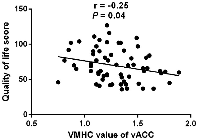
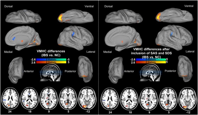
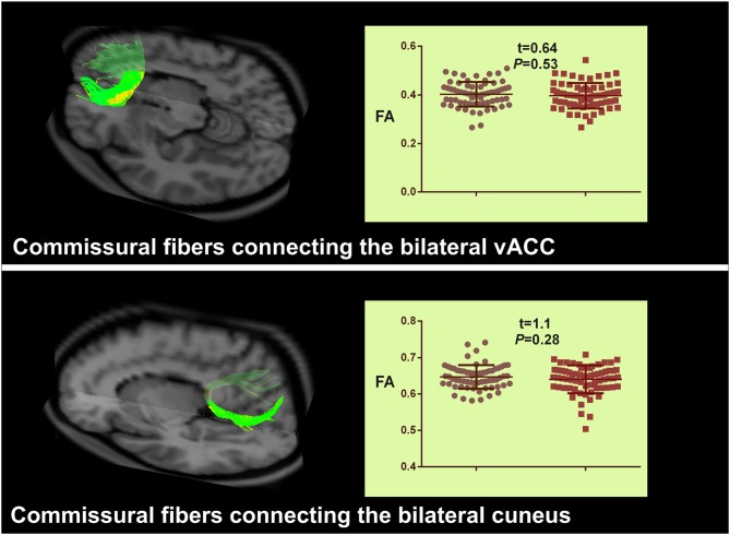
Neuroimaging studies have demonstrated that irritable bowel syndrome (IBS)-a relapsing functional bowel disorder-presents with disrupted brain connections. However, little is known about the alterations of interhemispheric functional connectivity and underlying structural connectivity in IBS. This study combined resting-state functional magnetic resonance imaging (rs-fMRI) and diffusion tensor imaging (DTI) to investigate changes in interhemispheric coordination in IBS patients. Resting-state functional and structural magnetic resonance images were acquired from 65 IBS patients and 67 healthy controls (HCs; matched for age, sex and educational level). Interhemispheric voxel-mirrored homotopic connectivity (VMHC) was calculated and compared between groups. Homotopic regions showing abnormal VMHC in patients were targeted as regions of interest (ROIs) for analysis of DTI tractography. The fractional anisotropy (FA), fiber number and fiber length were compared between groups. Statistical analysis was also performed by including anxiety and depression as covariates to evaluate their effect. A Pearson correlation analysis between abnormal interhemispheric connectivity and clinical indices of IBS patients was performed. Compared to HCs, IBS patients had higher interhemispheric functional connectivity between bilateral thalami, cuneus, posterior cingulate cortices (PCC), lingual gyri and inferior occipital/cerebellum lobes, as well as lower interhemispheric functional connectivity between bilateral ventral anterior cingulate cortices (vACC) and inferior parietal lobules (IPL). The inclusion of anxiety and depression as covariates abolished VMHC difference in vACC. Microstructural features of white matter tracts connecting functionally abnormal regions did not reveal any differences between the groups. VMHC values in vACC negatively correlated with the quality of life (QOL) scores of patients. In conclusion, this study provides preliminary evidence of the disrupted functional coordination rather than anatomic coordination between interhemispheric regions within the cortex-thalamus circuit in IBS patients, which could partly account for the enhanced visceral information processing and impaired endogenous pain or emotion inhibition associated with IBS.
神经影像学研究表明,肠易激综合征(IBS)——一种复发性功能性肠病——存在大脑连接中断的情况。然而,关于IBS患者半球间功能连接和潜在结构连接的改变知之甚少。本研究结合静息态功能磁共振成像(rs-fMRI)和扩散张量成像(DTI)来研究IBS患者半球间协调性的变化。从65例IBS患者和67名健康对照者(HCs;年龄、性别和教育水平相匹配)获取静息态功能和结构磁共振图像。计算并比较两组之间的半球间体素镜像同伦连接性(VMHC)。将患者中显示VMHC异常的同伦区域作为感兴趣区域(ROIs),用于分析DTI纤维束成像。比较两组之间的分数各向异性(FA)、纤维数量和纤维长度。还通过将焦虑和抑郁作为协变量进行统计分析,以评估它们的影响。对IBS患者异常半球间连接与临床指标进行Pearson相关分析。与HCs相比,IBS患者双侧丘脑、楔叶、后扣带回皮质(PCC)、舌回和枕叶/小脑下叶之间的半球间功能连接较高,而双侧腹侧前扣带回皮质(vACC)和顶下小叶(IPL)之间的半球间功能连接较低。将焦虑和抑郁作为协变量消除了vACC中VMHC的差异。连接功能异常区域的白质束的微观结构特征在两组之间未显示出任何差异。vACC中的VMHC值与患者的生活质量(QOL)评分呈负相关。总之,本研究提供了初步证据,表明IBS患者皮质-丘脑回路内半球间区域存在功能协调性而非解剖协调性中断,这可能部分解释了与IBS相关的内脏信息处理增强以及内源性疼痛或情绪抑制受损的原因。