Akarsu Cevher, Sahbaz Nuri Alper, Dural Ahmet Cem, Unsal Mustafa Gokhan, Kones Osman, Kocatas Ali, Halicioglu Ilkay, Alis Halil
General Surgery Department, University of Medical Sciences Bakirkoy Dr. Sadi Konuk Training and Research Hospital, Istanbul, Turkey.
JSLS. 2016 Oct-Dec;20(4). doi: 10.4293/JSLS.2016.00084.
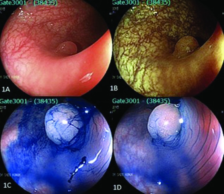
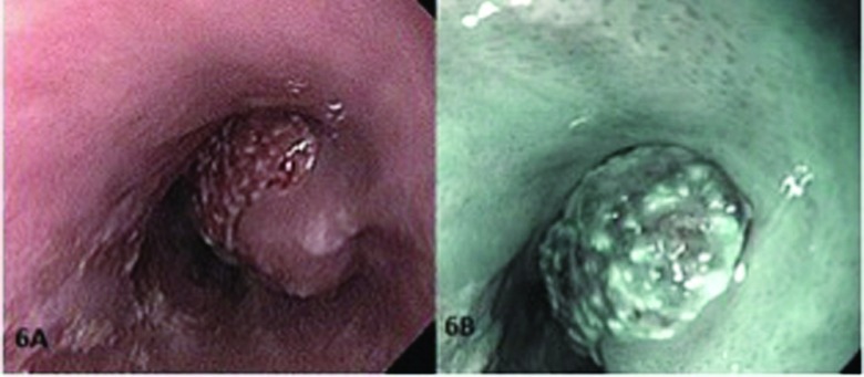
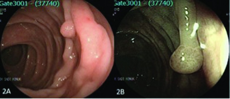
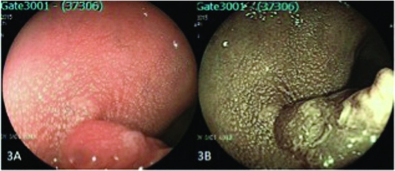
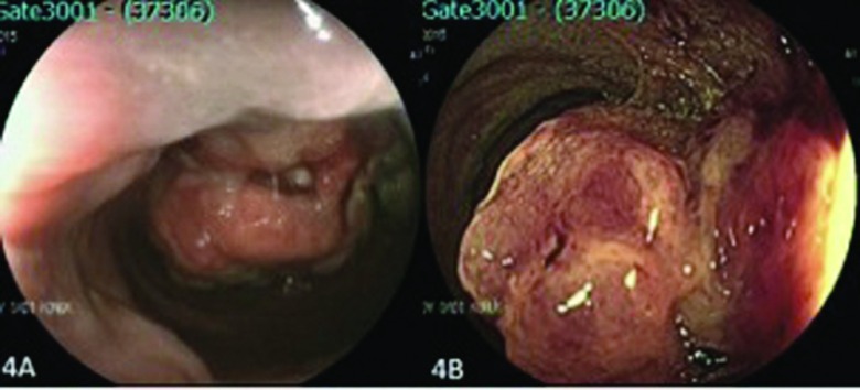
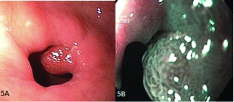
Gastrointestinal cancers are the most frequently occurring cancers worldwide. Diagnosis and removal of polyps during screening endoscopy decreases the prevalence of colon cancer and cancer-related mortality, and it is considered to be the gold standard in gastrointestinal system cancer prevention. Technological innovations in endoscopy have led to revolutionary developments in many areas. Flexible spectral imaging color enhancement (FICE) and narrow-band imaging (NBI) are forms of digital chromoendoscopy and enhance the endoscopic images without the need for a dye. This study seeks to evaluate the efficacy of FICE and NBI on polyp screening and real-time histologic diagnosis with endoscopy and to compare them.
A total of 134 patients (male/female = 72/62) and 161 polyps were evaluated with FICE or NBI, and real-time histologic diagnosis predictions were classified as neoplastic or nonneoplastic, according to Kudo's pit pattern classification. Pathological results and real-time endoscopic diagnoses were statistically interpreted for both FICE and NBI. Positive predictive value, negative predictive value, sensitivity, specificity, and accuracy rates were calculated and compared for both modalities.
When both systems were compared, the negative predictive value of NBI was found to be higher than that of FICE statistically ( < .001). Specificity and positive predictive value in the FICE group were higher than in the NBI group, but the difference was not statistically significant ( = .082 and = .153, respectively).
Aside from being safe in polyp detection, digital chromoendoscopy also helps the endoscopist in selecting the type of simultaneous intervention (eg, polypectomy, endomucosal resection, or submucosal dissection) by enabling endoscopic histologic diagnosis.
胃肠道癌是全球最常见的癌症。在筛查性内镜检查期间诊断并切除息肉可降低结肠癌的发病率及癌症相关死亡率,被视为胃肠系统癌症预防的金标准。内镜技术的创新已在许多领域带来了革命性的发展。灵活光谱成像色彩增强(FICE)和窄带成像(NBI)是数字染色内镜的形式,无需染料即可增强内镜图像。本研究旨在评估FICE和NBI在内镜息肉筛查及实时组织学诊断方面的效果,并对二者进行比较。
共134例患者(男/女 = 72/62)及161个息肉接受了FICE或NBI评估,并根据工藤的凹坑模式分类将实时组织学诊断预测分为肿瘤性或非肿瘤性。对FICE和NBI的病理结果及实时内镜诊断进行统计学分析。计算并比较两种方式的阳性预测值、阴性预测值、敏感性、特异性及准确率。
比较两种系统时,发现NBI的阴性预测值在统计学上高于FICE(<0.001)。FICE组的特异性和阳性预测值高于NBI组,但差异无统计学意义(分别为P = 0.082和P = 0.153)。
数字染色内镜除了在息肉检测中安全外,还通过实现内镜组织学诊断帮助内镜医师选择同步干预的类型(如息肉切除术、内镜黏膜切除术或黏膜下剥离术)。