Casquero-Veiga Marta, Hadar Ravit, Pascau Javier, Winter Christine, Desco Manuel, Soto-Montenegro María Luisa
CIBER de Salud Mental (CIBERSAM), Madrid, Spain.
Instituto de Investigación Sanitaria Gregorio Marañón, Madrid, Spain.
PLoS One. 2016 Dec 29;11(12):e0168689. doi: 10.1371/journal.pone.0168689. eCollection 2016.
To investigate metabolic changes in brain networks by deep brain stimulation (DBS) of the medial prefrontal cortex (mPFC), nucleus accumbens (NAcc) and dorsomedial thalamus (DM) using positron emission tomography (PET) in naïve rats.
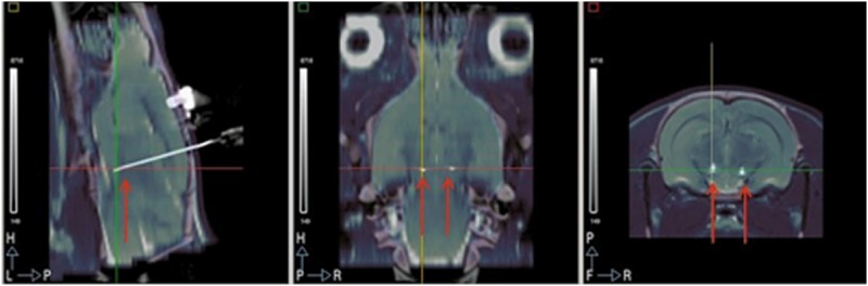
43 male Wistar rats underwent stereotactic surgery and concentric bipolar platinum-iridium electrodes were bilaterally implanted into one of the three brain sites. [18F]-fluoro-2-deoxy-glucose-PET (18FDG-PET) and computed tomography (CT) scans were performed at the 7th (without DBS) and 9th day (with DBS) after surgery. Stimulation period matched tracer uptake period. Images were acquired with a small-animal PET-CT scanner. Differences in glucose uptake between groups were assessed with Statistical Parametric Mapping.
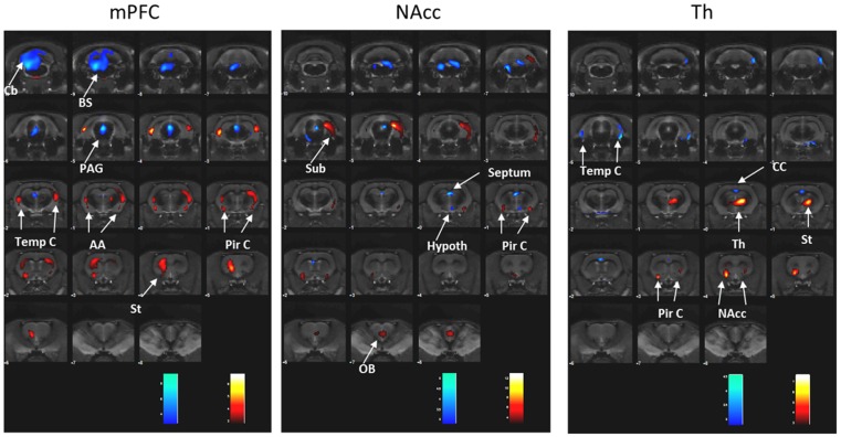
DBS induced site-specific metabolic changes, although a common increased metabolic activity in the piriform cortex was found for the three brain targets. mPFC-DBS increased metabolic activity in the striatum, temporal and amygdala, and reduced it in the cerebellum, brainstem (BS) and periaqueductal gray matter (PAG). NAcc-DBS increased metabolic activity in the subiculum and olfactory bulb, and decreased it in the BS, PAG, septum and hypothalamus. DM-DBS increased metabolic activity in the striatum, NAcc and thalamus and decreased it in the temporal and cingulate cortex.
DBS induced significant changes in 18FDG uptake in brain regions associated with the basal ganglia-thalamo-cortical circuitry. Stimulation of mPFC, NAcc and DM induced different patterns of 18FDG uptake despite interacting with the same circuitries. This may have important implications to DBS research suggesting individualized target selection according to specific neural modulatory requirements.
在未经处理的大鼠中,使用正电子发射断层扫描(PET)通过对内侧前额叶皮质(mPFC)、伏隔核(NAcc)和背内侧丘脑(DM)进行深部脑刺激(DBS)来研究脑网络中的代谢变化。
43只雄性Wistar大鼠接受立体定向手术,并将同心双极铂铱电极双侧植入三个脑区之一。在术后第7天(无DBS)和第9天(有DBS)进行[18F]-氟-2-脱氧葡萄糖-PET(18FDG-PET)和计算机断层扫描(CT)。刺激期与示踪剂摄取期匹配。使用小动物PET-CT扫描仪采集图像。用统计参数映射评估组间葡萄糖摄取的差异。
DBS诱导了位点特异性的代谢变化,尽管在三个脑靶点的梨状皮质中发现了共同的代谢活性增加。mPFC-DBS增加了纹状体、颞叶和杏仁核的代谢活性,并降低了小脑、脑干(BS)和导水管周围灰质(PAG)的代谢活性。NAcc-DBS增加了海马下托和嗅球的代谢活性,并降低了BS、PAG、隔区和下丘脑的代谢活性。DM-DBS增加了纹状体、NAcc和丘脑的代谢活性,并降低了颞叶和扣带回皮质的代谢活性。
DBS诱导了与基底神经节-丘脑-皮质回路相关的脑区18FDG摄取的显著变化。尽管mPFC、NAcc和DM的刺激与相同的回路相互作用,但它们诱导了不同模式的18FDG摄取。这可能对DBS研究具有重要意义,表明根据特定的神经调节需求进行个性化靶点选择。