Tu Sheng, Hu Fudong, Cai Wei, Xiao Liyan, Zhang Linlin, Zheng Hong, Jiang Qiong, Chen Lianglong
Department of Cardiology, Fujian Medical University Union Hospital, Fujian Institute of Coronary Heart Disease, Fuzhou, Fujian, People's Republic of China.
Department of Cardiology, The First Affiliated Hospital of Zhengzhou University, Zhengzhou, Henan, People's Republic of China.
Int J Cardiovasc Imaging. 2017 May;33(5):731-737. doi: 10.1007/s10554-016-1049-z. Epub 2016 Dec 30.
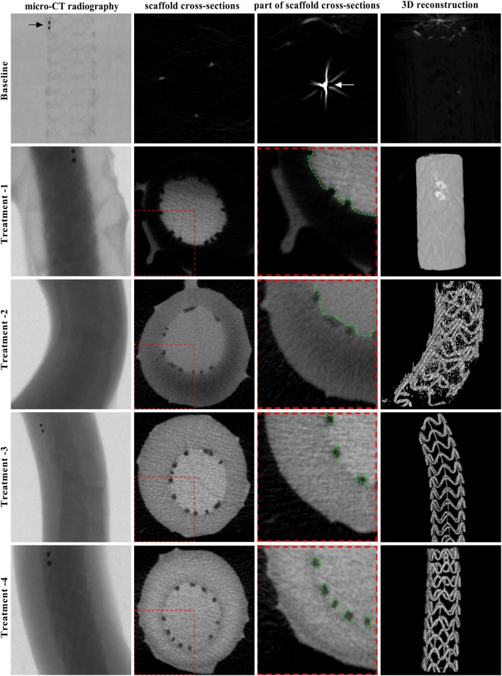
There are no previous studies showing how to visualize polymeric bioresorbable scaffolds (BRSs) by micro-computed tomography (mCT). There are no previous studies showing how to visualize polymeric bioresorbable scaffolds (BRSs) by micro-computed tomography (mCT). This study aimed to explore the feasibility of detecting polymeric BRS with 3-dimensional reconstruction of BRS images by contrast-enhanced mCT and to determine the optimal imaging settings. BRSs, made of poly-L-lactic acid (PLLA), were implanted in coronary bifurcation models. Five treatments were conducted to examine an optimal condition for imaging BRSs: Baseline treatment, samples were filled with normal saline and scanned with mCT immediately; Treatment-1, -2, -3 and -4, samples were filled with contrast medium and scanned with mCT immediately and 1, 2 and 3 h thereafter, corresponding to soaking time of contrast medium of 0, 1, 2 and 3 h. Compared to Baseline, mCT scanning completely discriminate the scaffold struts from the vascular lumen immediately after filling the samples with contrast agent but not from the vascular wall until the contrast agent soaking time was more than 2 h (Treatment-3 and -4). By setting 10-15 HU as a cut-point of CT values, the scaffold strut detectable rate at Baseline and Teatment-1, -2, -3 and -4 were 1.23 ± 0.31%, 1.65 ± 0.26%, 58.14 ± 12.84%, 97.97 ± 1.43% and 98.90 ± 0.38%, respectively (Treatment-3 vs. Treatment-2, p < 0.01); meanwhile, the success rate of 3D BRS reconstruction with high quality images at Baseline and Teatment-1, -2, -3 and -4 were 1.23%, 1.65%, 58.14%, 97.97% and 98.90%, respectively (Treatment-3 vs. Treatment-2, p < 0.01). In conclusions, reconstruction of 3D BRS images is technically feasible by contrast-enhanced mCT and soaking time of contrast agent for more than 2 h is necessary for complete separation of scaffold struts from the surrounding structures in the phantom samples.
以往没有研究表明如何通过微计算机断层扫描(mCT)对聚合物生物可吸收支架(BRS)进行可视化。以往没有研究表明如何通过微计算机断层扫描(mCT)对聚合物生物可吸收支架(BRS)进行可视化。本研究旨在探讨通过对比增强mCT对BRS图像进行三维重建来检测聚合物BRS的可行性,并确定最佳成像设置。将由聚-L-乳酸(PLLA)制成的BRS植入冠状动脉分叉模型中。进行了五种处理以检查BRS成像的最佳条件:基线处理,样品用生理盐水填充并立即用mCT扫描;处理-1、-2、-3和-4,样品用造影剂填充并立即以及之后1、2和3小时用mCT扫描,分别对应造影剂浸泡时间为0、1、2和3小时。与基线相比,在用造影剂填充样品后,mCT扫描能立即将支架支柱与血管腔完全区分开,但直到造影剂浸泡时间超过2小时(处理-3和-4)才能将其与血管壁区分开。通过将10 - 15 HU设置为CT值的截断点,基线和处理-1、-2、-3和-4时支架支柱的可检测率分别为1.23±0.31%、1.65±0.26%、58.14±12.84%、97.97±1.43%和98.90±0.38%(处理-3与处理-2相比,p<0.01);同时,基线和处理-1、-2、-3和-4时高质量图像三维BRS重建的成功率分别为1.23%、1.65%、58.14%、97.97%和98.90%(处理-3与处理-2相比,p<0.01)。总之,通过对比增强mCT对三维BRS图像进行重建在技术上是可行的,并且造影剂浸泡时间超过2小时对于在模型样品中将支架支柱与周围结构完全分离是必要的。