Byrne Aisling, Burke Christopher S, Keyes Tia E
School of Chemical Sciences , National Centre for Sensor Research , Dublin City University , Dublin 9 , Ireland . Email:
Chem Sci. 2016 Oct 19;7(10):6551-6562. doi: 10.1039/c6sc02588a. Epub 2016 Jun 30.
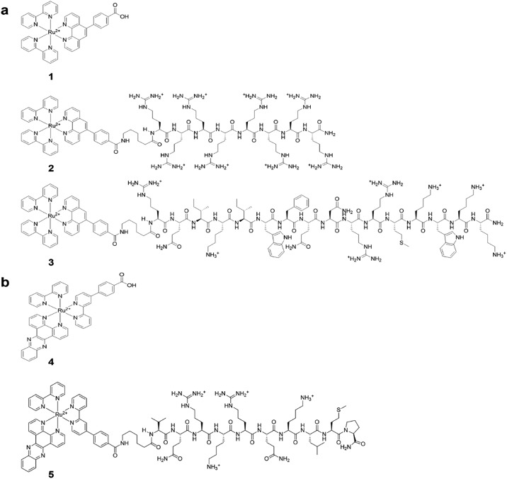
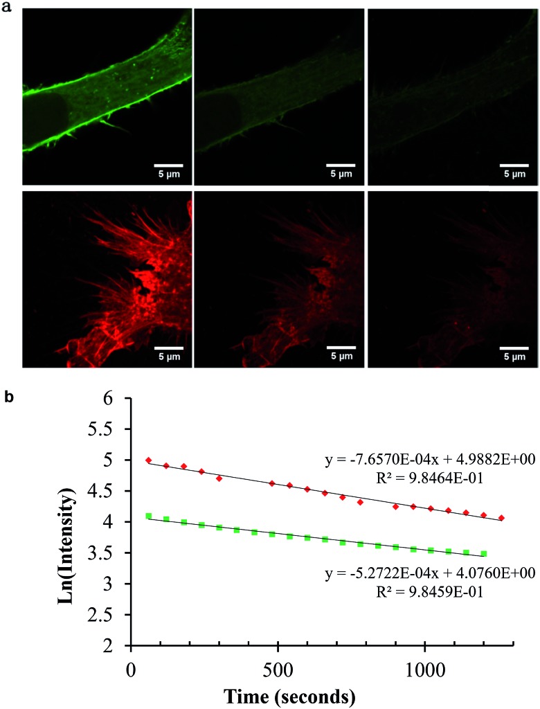
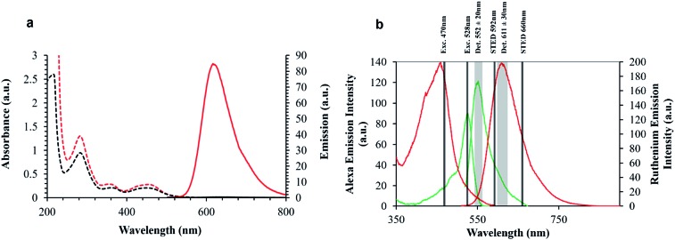
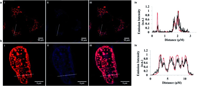
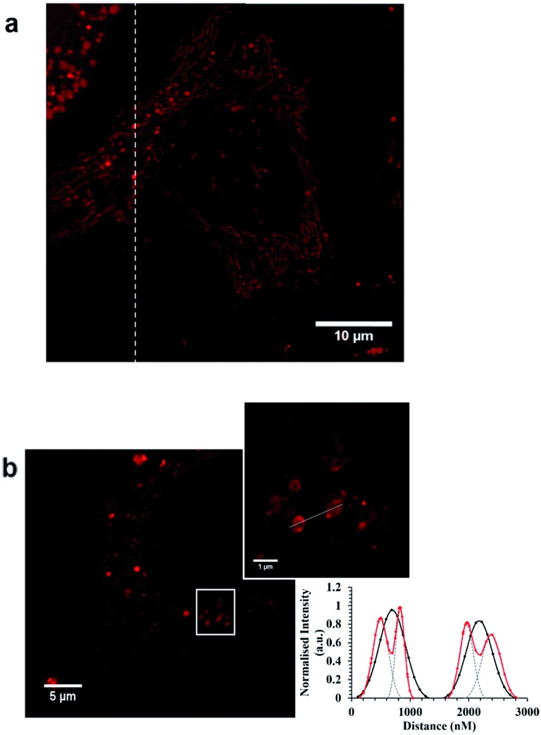
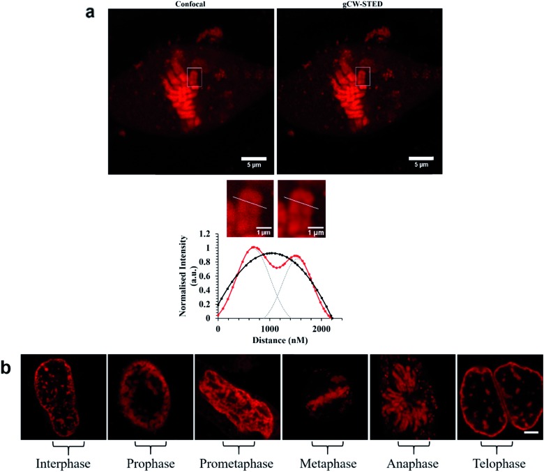
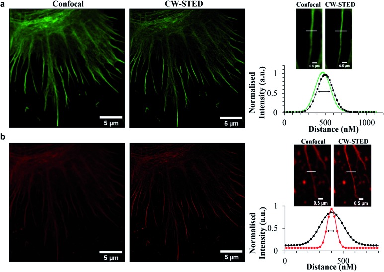
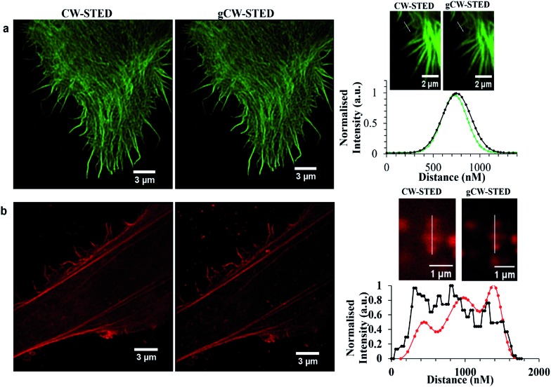
Fluorescence microscopy has undergone a dramatic evolution over the past two decades with development of super-resolution far-field microscopy methods that break the light diffraction limited resolution of conventional microscopy, offering unprecedented opportunity to interrogate cellular processes at the nanoscale. However, these methods make special demands of the luminescent agents used for contrast and development of probes suited to super-resolution fluorescent methods is still relatively in its infancy. In spite of their many photophysical advantages, metal complex luminophores have not yet been considered as probes in this regard, where to date, only organic fluorophores have been applied. Here, we report the first examples of metal complex luminophores applied as probes for use in stimulated emission depletion (STED) microscopy. Exemplified with endoplasmic reticulum and nuclear targeting complexes we demonstrate that luminescent Ru(ii) polypyridyl complexes can, through signal peptide targeting, be precisely and selectively delivered to key cell organelles without the need for membrane permeabilization, to give high quality STED images of these organelles. Detailed features of the tubular ER structure are revealed and in the case of the nuclear targeting probe we exploit the molecular light switch properties of a dipyrido[3,2-:2',3'-]phenazine containing complex which emits only on DNA/RNA binding to give outstanding STED contrast and resolution of the chromosomes within the nucleus. Comparing performance with a member of the AlexaFluor family commonly recommended for STED, we find that the performance of the ruthenium complexes is superior across both CW and gated STED microscopy methods in terms of image resolution and photostability. The large Stokes shifts of the Ru probes permit excellent matching of the stimulating depletion laser with their emission whilst avoiding anti-Stokes excitation. Their long lifetimes make them particularly amenable to gated STED, giving a much wider window for gating than traditional probes. Our findings indicate that ruthenium polypyridyl peptide targeted probes are a powerful new partner to STED microscopy, opening up new approaches to probe design for STED microscopy.
在过去二十年中,随着超分辨率远场显微镜方法的发展,荧光显微镜经历了巨大的变革。这些方法突破了传统显微镜受光衍射限制的分辨率,为在纳米尺度研究细胞过程提供了前所未有的机会。然而,这些方法对用于对比的发光剂有特殊要求,适用于超分辨率荧光方法的探针开发仍处于相对初期阶段。尽管金属络合物发光体具有许多光物理优势,但在这方面尚未被视为探针,迄今为止仅应用了有机荧光团。在此,我们报告了金属络合物发光体作为受激发射损耗(STED)显微镜探针的首个实例。以内质网和核靶向络合物为例,我们证明发光的钌(II)多吡啶络合物可通过信号肽靶向,在无需细胞膜通透化的情况下精确且选择性地递送至关键细胞器,从而获得这些细胞器的高质量STED图像。揭示了管状内质网结构的详细特征,对于核靶向探针,我们利用了含二吡啶并[3,2 - :2',3'-]吩嗪络合物的分子光开关特性,该络合物仅在与DNA/RNA结合时发光,从而在细胞核内提供出色的STED对比度和染色体分辨率。与通常推荐用于STED的AlexaFluor家族成员的性能进行比较,我们发现钌络合物在连续波(CW)和门控STED显微镜方法中的图像分辨率和光稳定性方面均表现更优。钌探针的大斯托克斯位移使得激发损耗激光与它们的发射能够完美匹配,同时避免反斯托克斯激发。它们的长寿命使其特别适用于门控STED,提供比传统探针宽得多的门控窗口。我们的研究结果表明,钌多吡啶肽靶向探针是STED显微镜强大的新伙伴,为STED显微镜的探针设计开辟了新途径。