• 文献检索
  • 文档翻译
  • 深度研究
  • 学术资讯
  • Suppr Zotero 插件Zotero 插件
  • 邀请有礼
  • 套餐&价格
  • 历史记录
应用&插件
Suppr Zotero 插件Zotero 插件浏览器插件Mac 客户端Windows 客户端微信小程序
定价
高级版会员购买积分包购买API积分包
服务
文献检索文档翻译深度研究API 文档MCP 服务
关于我们
关于 Suppr公司介绍联系我们用户协议隐私条款
关注我们

Suppr 超能文献

核心技术专利:CN118964589B侵权必究
粤ICP备2023148730 号-1Suppr @ 2026

文献检索

告别复杂PubMed语法,用中文像聊天一样搜索,搜遍4000万医学文献。AI智能推荐,让科研检索更轻松。

立即免费搜索

文件翻译

保留排版,准确专业,支持PDF/Word/PPT等文件格式,支持 12+语言互译。

免费翻译文档

深度研究

AI帮你快速写综述,25分钟生成高质量综述,智能提取关键信息,辅助科研写作。

立即免费体验

鼻孢子虫病:引起眼球突出的罕见病因及鼻窦肿物的影像学诊断难题

Rhinosporidiosis: A Rare Cause of Proptosis and an Imaging Dilemma for Sinonasal Masses.

作者信息

Dey Amit Kumar, Sharma Rajaram, Mittal Kartik, Kumar Puneeth, Murumkar Vivek, Mitkar Sumit, Hira Priya

机构信息

Department of Radiology, King Edward Memorial Hospital and Seth G.S. Medical College, Room No. 107, KEM Main Boy's Hostel, Parel, Mumbai 400012, India.

出版信息

Case Rep Otolaryngol. 2016;2016:3573512. doi: 10.1155/2016/3573512. Epub 2016 Nov 30.

DOI:10.1155/2016/3573512
PMID:28042489
原文链接:https://pmc.ncbi.nlm.nih.gov/articles/PMC5155101/
Abstract

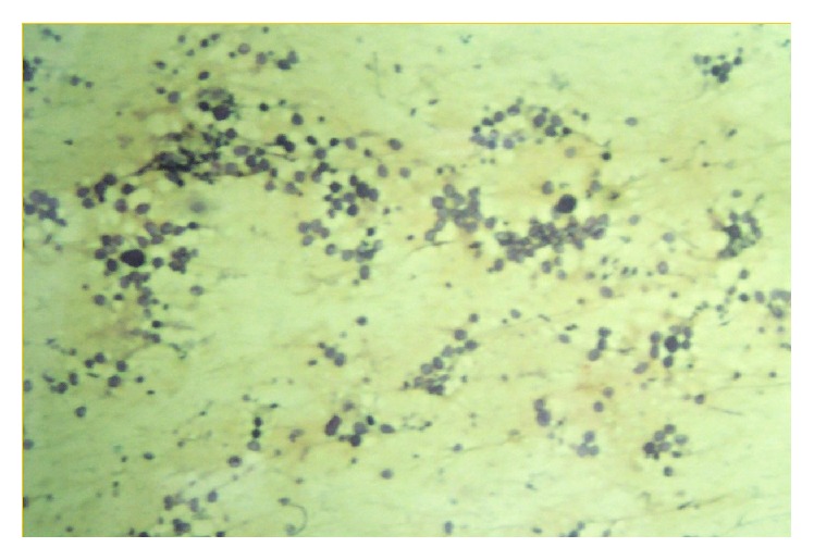
Rhinosporidiosis is a common disease entity in tropical countries; however, it can be encountered in other parts of the world as well due to increasing medical tourism. It may mimic other more malignant and vigorous pathologies of the involved part. We present a case of a 36-year-old male presenting with proptosis due to involvement of nasolacrimal duct which is rare. We will discuss typical CT and MRI features of the disease which were present in the case. For a surgeon and a radiologist, this is a necessary differential to be kept in mind for sinonasal masses. CT and MRI are invaluable investigations. However, FNAC is confirmatory. Both clinical and radiological aspects are required to reach correct diagnosis.

摘要

鼻孢子虫病在热带国家是一种常见的疾病实体;然而,由于医疗旅游的增加,在世界其他地区也可能遇到。它可能会模仿受累部位的其他更恶性和严重的病变。我们报告一例36岁男性因鼻泪管受累出现眼球突出的病例,这种情况较为罕见。我们将讨论该病例中出现的该病典型的CT和MRI特征。对于外科医生和放射科医生来说,这是鼻窦肿块需要牢记的一个必要鉴别诊断。CT和MRI是非常有价值的检查。然而,细针穿刺抽吸活检(FNAC)具有确诊意义。正确诊断需要临床和影像学两方面的信息。

https://cdn.ncbi.nlm.nih.gov/pmc/blobs/3bc3/5155101/51ead67394ec/CRIOT2016-3573512.003.jpg
https://cdn.ncbi.nlm.nih.gov/pmc/blobs/3bc3/5155101/6d6ca65bcc00/CRIOT2016-3573512.001.jpg
https://cdn.ncbi.nlm.nih.gov/pmc/blobs/3bc3/5155101/d931a1d6955e/CRIOT2016-3573512.002.jpg
https://cdn.ncbi.nlm.nih.gov/pmc/blobs/3bc3/5155101/51ead67394ec/CRIOT2016-3573512.003.jpg
https://cdn.ncbi.nlm.nih.gov/pmc/blobs/3bc3/5155101/6d6ca65bcc00/CRIOT2016-3573512.001.jpg
https://cdn.ncbi.nlm.nih.gov/pmc/blobs/3bc3/5155101/d931a1d6955e/CRIOT2016-3573512.002.jpg
https://cdn.ncbi.nlm.nih.gov/pmc/blobs/3bc3/5155101/51ead67394ec/CRIOT2016-3573512.003.jpg

相似文献

1
Rhinosporidiosis: A Rare Cause of Proptosis and an Imaging Dilemma for Sinonasal Masses.鼻孢子虫病:引起眼球突出的罕见病因及鼻窦肿物的影像学诊断难题
Case Rep Otolaryngol. 2016;2016:3573512. doi: 10.1155/2016/3573512. Epub 2016 Nov 30.
2
Lacrimal Sac Rhinosporidiosis and Surgical Management by Transnasal Endoscopic Excision: A Case Series.泪囊鼻孢子虫病及经鼻内镜切除的外科治疗:病例系列
Laryngoscope. 2018 Dec;128(12):2693-2696. doi: 10.1002/lary.27322. Epub 2018 Sep 8.
3
Magnetic Resonance Imaging in Evaluation of Sinonasal Masses with Histopathological Correlation.磁共振成像在鼻窦肿物评估中的应用及其与组织病理学的相关性
Mymensingh Med J. 2018 Jan;27(1):26-33.
4
Imaging in Rare and Atypical Sinonasal Masses: An Interesting Case Series.罕见及非典型鼻窦肿物的影像学表现:一组有趣的病例系列
J Clin Diagn Res. 2015 Dec;9(12):TR01-5. doi: 10.7860/JCDR/2015/14994.6900. Epub 2015 Dec 1.
5
Infant with nasolacrimal sinonasal myxoma: Diffusion MRI features.患有鼻泪管鼻窦黏液瘤的婴儿:扩散加权磁共振成像特征
Radiol Case Rep. 2016 Feb 17;10(2):1104. doi: 10.2484/rcr.v10i2.1104. eCollection 2015.
6
Isolated Primary Rhinosporidiosis of the Parotid Duct: A Rare Presentation.腮腺导管孤立性原发性鼻孢子虫病:一种罕见的表现。
Iran J Otorhinolaryngol. 2020 May;32(110):193-196. doi: 10.22038/ijorl.2020.43051.2408.
7
Disseminated rhinosporidiosis.播散性鼻孢子虫病。
J Laryngol Otol. 2013 Oct;127(10):1020-4. doi: 10.1017/S0022215113002193. Epub 2013 Oct 14.
8
Educational report: A case of lacrimal sac rhinosporidiosis.教育报告:一例泪囊鼻孢子虫病病例。
Orbit. 2016 Oct;35(5):254-7. doi: 10.1080/01676830.2016.1193529. Epub 2016 Aug 10.
9
Benign sinonasal masses: a clinicopathological and radiological profile.鼻腔鼻窦良性肿物:临床病理与影像学特征
Kathmandu Univ Med J (KUMJ). 2013 Jan-Mar;11(41):4-8. doi: 10.3126/kumj.v11i1.11015.
10
A Rare Presentation of Rhinosporidiosis.鼻孢子虫病的罕见表现
Indian J Otolaryngol Head Neck Surg. 2017 Dec;69(4):559-562. doi: 10.1007/s12070-017-1080-y. Epub 2017 Feb 7.

引用本文的文献

1
Nasal rhinosporidiosis masquerading as pyogenic granuloma in a pediatric patient at a Zonal Referral Hospital in Central Tanzania: Case report.坦桑尼亚中部一家地区转诊医院里,一名儿科患者身上出现伪装成化脓性肉芽肿的鼻孢子虫病:病例报告。
Clin Case Rep. 2022 Jun 19;10(6):e5977. doi: 10.1002/ccr3.5977. eCollection 2022 Jun.

本文引用的文献

1
Imaging features of rhinosporidiosis on contrast CT.增强CT上鼻孢子虫病的影像学特征。
Indian J Radiol Imaging. 2013 Jul;23(3):212-8. doi: 10.4103/0971-3026.120267.
2
Rhinosporidiosis affecting multiple parts of the body.鼻孢子虫病累及身体多个部位。
Trop Doct. 2012 Jul;42(3):174-5. doi: 10.1258/td.2012.120035.
3
Bronchial rhinosporidiosis: An unusual presentation.支气管鼻孢子虫病:一种不寻常的表现。
Lung India. 2012 Apr;29(2):173-5. doi: 10.4103/0970-2113.95336.
4
Nasal rhinosporidiosis.鼻孢子虫病
J Natl Med Assoc. 2008 Jun;100(6):713-5. doi: 10.1016/s0027-9684(15)31347-x.
5
Disseminated cutaneous rhinosporidiosis.播散性皮肤鼻孢子虫病。
Dermatol Online J. 2005 Mar 1;11(1):19.
6
Nasal rhinosporidiosis; report of a case in natal.鼻孢子虫病;纳塔尔省一例报告。
S Afr Med J. 1951 Apr 21;25(16):270-1.
7
Generalized rhinosporidiosis with visceral involvement; report of a case.伴有内脏受累的播散性鼻孢子虫病;病例报告
AMA Arch Derm. 1959 Jul;80(1):22-6. doi: 10.1001/archderm.1959.01560190024003.
8
Oculosporidiosis in eastern Nepal: a report of five cases.尼泊尔东部的眼孢子虫病:5例报告
Southeast Asian J Trop Med Public Health. 2002 Jun;33(2):362-4.
9
A verrucous lesion of the palm.手掌的疣状病变。
Postgrad Med J. 2002 May;78(919):302, 305-6. doi: 10.1136/pmj.78.919.302.
10
Disseminated cutaneous rhinosporidiosis.
J Dermatol. 1998 Aug;25(8):527-32. doi: 10.1111/j.1346-8138.1998.tb02449.x.